Learn How Education And Movement Can Help Frozen Shoulder

If I’ve heard it once, I’ve heard it 1000 times in my career (okay this is an exaggeration, it’s probably only a few dozen times), “I think my frozen shoulder is acting up!”

Meanwhile, the patient is moving their arm all over and wincing with a bit of pain.

The very name is enough to strike long term fear into a patient.

“Frozen Shoulder” it just sounds like something utterly debilitating, no wonder patients catastrophize over the diagnosis.

Part of the problem with this diagnosis is that all too often as soon as someone has an issue with their shoulder, it gets labeled, labeled with a damning diagnosis, and usually an improper one.

So, how do we convince someone their shoulder is probably okay and just needs to be de-sensitized, or actually realize frozen shoulder is the issue? Well, there are several ways, and while we can’t “diagnose” there are certainly some signs and symptoms we can look at which will help us recognize the difference and refer out when necessary.

Getting To Know Frozen Shoulder

Most of the time a detailed intake form, case history, and clinical examination should be enough to manage those who are coming in with shoulder pain, however, there are things we need to look for during this to determine the severity of a shoulder issue.

Frozen shoulder’s clinical name is “adhesive capsulitis” and is characterized by patients experiencing pain along with limited range of motion and disability of the glenohumeral joint which lasts anywhere from 1-24 months. There are two types of adhesive capsulitis:

  1. Idiopathic (primary): occurs spontaneously from a chronic inflammatory response (possibly an abnormal immune system response).
  2. Secondary adhesive capsulitis: happens after a shoulder injury or surgery and can be associated with conditions like diabetes, rotator cuff injury, cerebrovascular accident, or cardiovascular disease.

It seems those with diabetes (10-36% of diabetic patients) are quite prone to dealing with frozen shoulder as both types I and type II diabetics are susceptible and have worse outcomes compared to non-diabetics. They also experience more severe symptoms and are more resistant to treatment. Those who have had a stroke are also quite susceptible as it happens to 25% of stroke patients within 6 months, which is likely due to some muscle spasticity on the affected side. Some studies have also shown an association with Dupuytren’s disease, hypothyroidism, and Parkinson’s disease (however these last few are much rarer).

Mostly this affects people in their 50’s with the peak age being 56, but rarely happening to people under 40, and more commonly affects women than men (sorry ladies). However, some research suggests a high prevalence of shoulder issues among the elderly, who aren’t seeking medical attention for the issue.

One study tried to develop a new clinical sign to help diagnose a frozen shoulder with something called the “Coracoid Pain Test”. This is essentially putting digital pressure on the coracoid process, which creates more intense pain compared to the unaffected shoulder. In the study, 96.4% of patients with this condition complained of pain when the test was done, which they argue shows a high specificity rating, but I don’t know if this is widely accepted yet.

If we have a patient come in who is experiencing shoulder pain, trying to get a differential diagnosis is important (even though we can’t diagnose), but if we look at the above instances, these are all things which could be on our intake form that could help us narrow down and understand that maybe our patient is dealing with frozen shoulder as opposed to just some simple shoulder pain. One other thing that stands out as being consistent with a frozen shoulder is the complete loss of external rotation.

Once we understand this, it is also important to understand the “phases” this condition goes through.

There are 3 phases with varying degrees of length:

  1. The painful phase:
    • Pain with AROM & PROM.
    • Reduced flexion, abduction, and rotation.
    • Pain worse at night.
    • Duration lasting 10-36 weeks.
  2. Adhesive “frozen” phase:
    • Pain starts to subside (still bad at full range) but still stiff.
    • Almost no external rotation.
    • Rigid “end feel”.
    • Duration 9-15 months.
  3. Resolution “thawing” phase:
    • Spontaneous improvement in ROM.
    • Minimal pain.
    • Happens during 15-24 months since issues started.

While it is quite common for the symptoms to resolve themselves, it does take a considerable amount of time for that to happen and of course, this depends on whether things like diabetes are influencing the healing process. There are studies showing that 39% of people had a full recovery, the remaining 61% had some issues with pain and or range of motion.

However, there are things we can do to help move this along and education with movement looks like the primary treatment.

Of course, the treatment we are giving must be tailored not only to the patient but also the phase of the condition.

During the painful phase, the main thing we are looking for is pain relief and movement within pain-free tolerances and using graded exposure to get to the edges of painful movement. We did an article a couple of weeks ago where you can see how to do this by clicking HERE. While much of the literature points to the use of NSAIDs, there isn’t a lot to confirm its effectiveness for frozen shoulder.

One study on Idiopathic Adhesive Capsulitis showed good success with an exercise program that involved a four-direction shoulder stretching program that included passive forward flexion, passive external rotation, passive horizontal adduction, and passive internal rotation. With this program they had 64% of patients report a satisfactory outcome, 7% not satisfied, and 5% who went for surgery.

Treatments during the adhesive phase should be more aggressive toward longer stretches and a low load to push toward an increase in range of motion.

When these exercise interventions don’t work, the patient is often referred for surgery or for manipulation under anesthesia and have relatively good outcomes. There is also well-documented use of injected steroids, which when combined with manual therapy have some good outcomes as well. Although, in reading over Paul Ingraham’s post on frozen shoulder, there is also a risk of causing shoulder issues with various types of injections, so this should be considered as well.

Much of this is going to be left up to your clinical decision making, but having a good understanding of the timelines and what is happening, along with feedback from your patient should give you a good idea of how to manage this, should someone come in for treatment.

Educating

As mentioned in this post, education is a major part of helping someone with this condition. 

Unfortunately, this probably isn’t done as much as it should be. While most patients are probably looking for a “quick fix”, the reality is, this is just going to take some time and effort on their part and yours. 

There is a normal course the condition takes and at the 12-24 month period it falls into a resolution phase and there is a greater improvement in range of motion. While any patient would look for complete resolution one study showed at the 5-10 year follow up of 41 patients: 

  • 39% had full recovery.
  • 54% had some limitation without functional disability.
  • 7% had functional limitations. 

And still another study showed 50% of their patients had some degree of pain and stiffness seven years after the condition started. 

However, the above studies did show that the longer the person was in the stiffness stage, the longer the recovery stage, there was a direct correlation. So, perhaps proper education and movement in the painful phase could, in turn, shorten the stiffness and recovery stage?

Interestingly one of the previously mentioned studies showed that prior “physical therapy treatment and a workman’s compensation claim or pending litigation were the only variables that were associated with the eventual need for manipulation or capsular release”.

This is a fact I find really interesting. While some argue that the biopsychosocial approach to pain isn’t in our scope, how can we look at that study and say these other factors are not a contributing factor to a persons pain and disability? In my old job I was told by a compensation representative that their studies had shown if someone was off work for 18 months on an injury claim, chances are they were never going back to that job. And here we have studies showing us that a compensation claim is one of the contributing factors to needing more aggressive treatment for this condition. While we cannot counsel a patient on this, it is something we should be cognizant of when treating them (if a compensation claim is part of their issue).

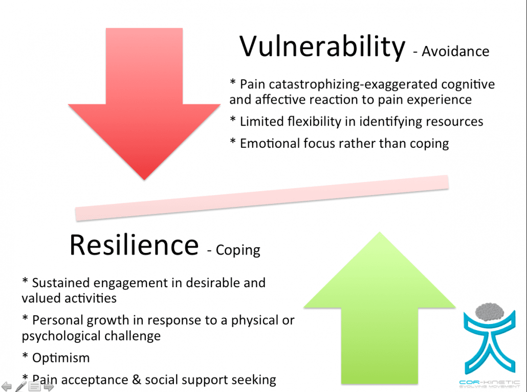
While we would never want to tell a patient there is only a 50% chance that after seven years they would be pain-free, we do want to try to educate, encourage, and build resilience with them through each phase of this condition, giving them hope for the most positive outcome possible. Reassuring them that there is a bit of a longer recovery process compared to other shoulder issues, but that full recovery is possible will probably bring a better chance of shortening the stiffness, and recovery stages.

Many Orthopedic Surgeries Don’t Work Better Than Placebo

Unlike drugs, orthopedic surgeries can be sold to the public before they undergo rigorous testing to ensure they are safe and effective. Thus, millions of surgeries for knee, shoulder, and back pain have been done without studies to confirm they actually work. Recently, research has found that many popular surgeries (but not all) work no better than a placebo. And yet these surgeries are still done at the rate of hundreds of thousands per year. This is something you should know if you are considering surgery, or in the business of treating chronic pain.

Knee Surgery

Osteoarthritis of the knee is common but does not necessarily result in pain — many people have arthritis and no pain at all. But surgery to correct arthritis is popular, numbering as many as a half-million per year in the U.S. alone. (1)

About 15 years ago, two of the most common procedures were debridement (removal of damaged cartilage or bone) or lavage (irrigation with saline solution). The goal of either surgery was removal of rough fragments of cartilage that may be irritating the joint. The surgery’s benefits were known to be not so impressive, so people started to wonder whether they were caused by placebo, or could be achieved with less invasive treatment.

To answer these questions, a study was done using a “sham” surgery: one group of patients received real knee surgery, and the other a fake, which involved just an incision on the skin. At several times over the course of a couple of years, the two groups reported their levels of knee pain and function. The result? The sham group did just as well as the surgical group at all points in time. (2) This strongly suggests the surgery worked by changing psychology, not structure.

This study was slow to have an effect on the behavior of surgeons. Years later, hundreds of thousands of these surgeries were still being performed at the cost of $3 billion a year. (3) Subsequent research confirmed they provide no more benefit than common sense interventions like exercise, weight loss, and the occasional use of over-the-counter pain medicine. (4) Debridement and lavage eventually became less common but were soon replaced by arthroscopic partial meniscectomy. But the new surgery proved to be equally weak. Like its predecessors, it worked no better than a sham.

In 2015, researchers summarized the results of nine studies on arthroscopic procedures for the knee and concluded that they showed little if any benefit, and the possibility for serious harm. (5) Further studies in 2017 came to similar conclusions. (5a)

A recent editorial in the British Journal of Medicine offered the scathing opinion that arthroscopic surgery for knee pain is: “a highly questionable practice without supporting evidence of even moderate quality.” (6) A clinical guideline issued in 2017 made a “strong recommendation against the use of arthroscopy in nearly all patients with degenerative knee disease” and noted that “further research is unlikely to alter this recommendation.” (7) In spite of this advice, arthroscopic knee surgery continues to be the most common orthopedic procedure in the U.S., with close to 700,000 performed each year. (6)

Why the disconnect between research and practice? The simple answer is that many doctors trust their personal experience over-controlled research. They will say they have seen surgery provide benefit after conservative therapies failed. Surely this sometimes occurs, but we should be skeptical that the cause is structural change in the knee. Instead, successful treatment for chronic knee pain, through surgery or otherwise, may be caused by complex changes in psychological or neurological processes.

Back Surgery

As with knees, there is a wealth of evidence that backs can have major structural damage and still not hurt. Further, backs can hurt a lot even when they have no apparent damage. In fact, this is quite common — most chronic low back pain is classified as “non-specific”, meaning it cannot be explained in reference to a mechanical or structural cause. Although there is only a poor correlation between back pain and structural back damage, there are many surgeries to correct any damage that happens to appear on an MRI. Several perform no better than conservative care.

For example, vertebroplasty attempts to treat back pain by injecting bone cement into fractured vertebrae. A study published in 2003 showed it works as well as no treatment at all after six weeks. (8) In 2009, two studies showed it worked no better than a sham procedure for fractures related to osteoporosis. (9)

Low back fusion is a common procedure and is intended to enhance low back stability. In 2013, the Spine Journal published a study comparing spinal fusion to non-operative treatments like cognitive behavioral therapy and exercise. It found no difference in long term outcomes and concluded that “the use of lumbar fusion in chronic low back pain patients should not be favored” over conservative care and exercise. (10)

In 2014, an analysis of multiple studies concluded that:

There is strong evidence that lumbar fusion is not more effective than conservative treatment in reducing perceived disability because of chronic low back pain among patients with degenerative spinal diseases. It is unlikely that further research on the subject would considerably affect this conclusion.(11)

As with knee surgery, the research showing the relative ineffectiveness of surgery has been slow to affect practice. Nikolai Bogduk, an internationally renowned anatomist and back pain expert, explains that:

Surgeons and others believe that surgery is effective for back pain. They base this belief either on their own experience or on observational studies. This belief is, by and large, not vindicated by the outcomes in well-reported clinical trials. Those trials indicate that only a small proportion of patients do well from surgery.(12)

Shoulder Surgery

The research on shoulder surgery is less clear, but recent studies reveal a similar pattern. MRIs consistently show that a great many people without pain have torn rotator cuffs. Despite this fact, the most common diagnoses to explain shoulder pain is “impingement” on the rotator cuff by the acromion process.

Surgeries to correct these conditions include rotator cuff repair, or acromioplasty, where a portion of the acromion is removed. In the United States, there are almost half a million of these surgeries performed each year, most of them on shoulders where the damage is degenerative as opposed to traumatic. (13)

Although we cannot rule out the possibility that surgery will be substantially more effective than exercise for certain patients (14), several studies have found that popular shoulder surgeries, such as acromioplasty, are no better than exercise. (15) Further, it has been shown that surgeries for labral repair and biceps tendinosis cannot outperform sham surgery. (16)

There is also evidence suggesting that when surgery is effective at relieving pain, it is not because of structural repair. (17) MRIs of repaired rotator cuffs taken within a year after surgery often show that the cuff has fallen apart again, even though the patient has recovered. According to Dr. Lawrence Gullotta, “When your rotator cuff is torn, you attribute all your pain and dysfunction to your torn rotator cuff, then you have it fixed and you feel better, but sometimes when you take an ultrasound or an MRI, the rotator cuff looks exactly like it did before you had the surgery.” (18)

A 2017 study compared two kinds of shoulder arthroscopic surgery (with and without decompression), and found that neither provided a clinically significant benefit over no treatment at all. (19) An editorial said these findings “send a strong message that the burden of proof now rests on those who wish to defend the standpoint that shoulder arthroscopy is more effective than non-surgical interventions.” (20)

A blog post in the British Journal of Sports Medicine summed up the situation as follows: “in the light of the current evidence base, the benefits of surgical intervention for the treatment of [shoulder pain] seem glorified and overrated.” (21)

Conclusion

Let me make something clear: None of the above means we should not trust orthopedic surgeons, or that surgery should never be used to treat chronic pain. Many surgeries have been proven to work and are definitely a good idea under certain circumstances. I personally know many people who have received excellent and ethical advice from their surgeons, and dramatic benefits from surgery. By contrast, I also know people who got a surgery that had been proven to be ineffective, were never advised of the relevant research, and ultimately did not get a good result.

I think the lesson here is that the medical establishment has some biases in the way it treats chronic pain. It favors treatments based on simple structural explanations for pain and tends to ignore complex neurophysiological processes that may be more important. We need to be aware of these biases and learn as much as possible about the complexity of pain so that treatment can be improved. And chronic pain clients need to ask their surgeons some good questions before going under the knife.


The above article is an adapted excerpt from my new book, Playing With Movement: How to Explore the Many Dimensions of Physical Health and Performance


References

1. Heidari, Behzad. 2011. “Knee Osteoarthritis Prevalence, Risk Factors, Pathogenesis and Features: Part I.” Caspian Journal of Internal Medicine 2 (2): 205–12.2.

2. Lubowitz, James H. 2002. “A Controlled Trial of Arthroscopic Surgery for Osteoarthritis of the Knee.” Arthroscopy 18 (8): 950–51. http://www.ncbi.nlm.nih.gov/pubmed/12368798.

3. Why ‘Useless’ Surgery Is Still Popular. http://www.nytimes.com/2016/08/04/upshot/the-right-to-know-that-an-operation-is-next-to-useless.html?_r=2

4. Kirkely, Birmingham, et al. 2008. “A Randomized Trial of Arthroscopic Surgery for Osteoarthritis of the Knee.” New England Journal. Vol. 359, 1097.

5. Thorlund, J. B., C. B. Juhl, E. M. Roos, and L. S. Lohmander. 2015. “Arthroscopic Surgery for Degenerative Knee: Systematic Review and Meta-Analysis of Benefits and Harms.” Bmj 350 (jun16 3): h2747–h2747. doi:10.1136/bmj.h2747.

5a. “Brignardello-Petersen et al. (2017). Knee Arthroscopy Versus Conservative Management in Patients with Degenerative Knee Disease: A Systematic Review. BMJ Open 7(5), e016114; Sihvonen et al. (2017). Arthroscopic Partial Meniscectomy Versus Placebo Surgery for a Degenerative Meniscus Tear: A 2-Year Follow-Up of the Randomised Controlled Trial. Annals of the Rheumatic Diseases. 77(2), 188–195.”

6. Järvinen, Teppo L N, and Gordon H Guyatt. 2016. “Arthroscopic Surgery for Knee Pain.” BMJ (Clinical Research Ed.) 354 (July). British Medical Journal Publishing Group: i3934. doi:10.1136/BMJ.I3934.

7. Siemieniuk, Reed A C, Ian A Harris, Thomas Agoritsas, Rudolf W Poolman, Romina Brignardello-Petersen, Stijn Van de Velde, Rachelle Buchbinder, et al. 2017. “Arthroscopic Surgery for Degenerative Knee Arthritis and Meniscal Tears: A Clinical Practice Guideline.” Bmj, j1982. doi:10.1136/bmj.j1982.

8. Diamond, Terrence H, Bernard Champion, and William A Clark. 2003. “Management of Acute Osteoporotic Vertebral Fractures: A Nonrandomized Trial Comparing Percutaneous Vertebroplasty with Conservative Therapy.” The American Journal of Medicine 114 (4): 257–65. doi:10.1016/S0002–9343(02)01524–3.

9. Kallmes, David F., Bryan A. Comstock, Patrick J. Heagerty, Judith A. Turner, David J. Wilson, Terry H. Diamond, Richard Edwards, et al. 2009. “A Randomized Trial of Vertebroplasty for Osteoporotic Spinal Fractures.” New England Journal of Medicine 361 (6): 569–79. doi:10.1056/NEJMoa0900563.

10. Mannion, Anne F., Jens Ivar Brox, and Jeremy C.T. Fairbank. 2013. “Comparison of Spinal Fusion and Nonoperative Treatment in Patients with Chronic Low Back Pain: Long-Term Follow-up of Three Randomized Controlled Trials.” The Spine Journal 13 (11): 1438–48. doi:10.1016/j.spinee.2013.06.101.

11. Saltychev, Mikhail, Merja Eskola, and Katri Laimi. 2014. “Lumbar Fusion Compared with Conservative Treatment in Patients with Chronic Low Back Pain.” International Journal of Rehabilitation Research 37 (1): 2–8. doi:10.1097/MRR.0b013e328363ba4b.

12. Bogduk, Nikolai, and Gunnar Andersson. 2009. “Is Spinal Surgery Effective for Back Pain?” F1000 Medicine Reports 1 (July): 27–29. doi:10.3410/M1–60.

13. Jain, Nitin B, Laurence D Higgins, Elena Losina, Jamie Collins, Philip E Blazar, and Jeffrey N Katz. 2014. “Epidemiology of Musculoskeletal Upper Extremity Ambulatory Surgery in the United States.” BMC Musculoskeletal Disorders 15 (1): 4. doi:10.1186/1471–2474–15–4.

14. Steuri, Ruedi, Martin Sattelmayer, Simone Elsig, Chloé Kolly, Amir Tal, Jan Taeymans, and Roger Hilfiker. 2017. “Effectiveness of Conservative Interventions Including Exercise, Manual Therapy and Medical Management in Adults with Shoulder Impingement: A Systematic Review and Meta-Analysis of RCTs.” British Journal of Sports Medicine, bjsports-2016–096515. doi:10.1136/bjsports-2016–096515.

15. Ketola, S., J. Lehtinen, T. Rousi, M. Nissinen, H. Huhtala, Y. T. Konttinen, and I. Arnala. 2013. “No Evidence of Long-Term Benefits of Arthroscopicacromioplasty in the Treatment of Shoulder Impingement Syndrome: Five-Year Results of a Randomised Controlled Trial.” Bone and Joint Research 2 (7): 132–39. doi:10.1302/2046–3758.27.2000163.

16. Brox, Jens Ivar, Cecilie Piene Schrøder, Øystein Skare, Petter Mowinckel, and Olav Reikerås. 2017. “Author Response — sham Surgery versus Labral Repair or Biceps Tenodesis for Type II SLAP Lesions of the Shoulder: A Three-Armed Randomised Clinical Trial.” British Journal of Sports Medicine, bjsports-2017–098251. doi:10.1136/bjsports-2017–098251.

17. McElvany, Matthew D., Erik McGoldrick, Albert O. Gee, Moni Blazej Neradilek, and Frederick A. Matsen. 2015. “Rotator Cuff Repair.” The American Journal of Sports Medicine 43 (2). SAGE PublicationsSage CA: Los Angeles, CA: 491–500. doi:10.1177/0363546514529644.

18. Large Study of Arthroscopic Rotator Cuff Repair Reveals Some Surprises. http://www.hss.edu/newsroom_study-arthroscopic-rotator-cuff-repair-surprises.asp

19. Beard et al. (2017). Arthroscopic Subacromial Decompression for Subacromial Shoulder Pain (Csaw): A Multicentre, Pragmatic, Parallel Group, Placebo-Controlled, Three-Group, Randomised Surgical Trial. The Lancet. 391, 329–38.

20. Schreurs et al. (2017). No Benefit of Arthroscopy in Subacromial Shoulder Pain. The Lancet. 6736 (17).21. “Unnecessary Shoulder Surgery on the Rise — BJSM Blog — Social Media’s Leading SEM Voice.” http://blogs.bmj.com/bjsm/2015/01/06/the-sexy-scalpel-unnecessary-shoulder-surgery-on-the-rise/.

Resilience – What It Is And Why Is It A Big Deal For Recovery?


Resilience is a term used more and more by therapists, some might suggest it is a buzz word, so I wanted to put down my thoughts about this complex subject. I have wanted to write this blog for a while and the struggle I have had here is to keep it bloggy, not too academic and above all not too long! I have tried to strike a balance between research and also some personal experiences of resilience I have collected.

Before we get into it, there are 3 main points I would like to make in this blog.

Firstly, resilience is NOT about manning (or womaning) up, it is NOT SIMPLY about just keeping going. It’s a flexible, adaptable state that recognizes the importance of specific actions, mental or physical, that have a positive impact on life.

Secondly, resilience is not a binary state between resilience and not resilient. It is a continuum that we move along dependent on a balance of internal and external factors.

Thirdly that resilience looks and feels different between people. What you may regard as resilience MAY NOT be the same as the person you are working with.

What Is Resilience?

Resilience is defined by Sturgeon *HERE* as,

“Maintenance of positive physical and emotional functioning in spite of significant difficulty or challenge”

So resilience is both a physical and a psychological thing. It is important that we don’t separate the two, however (much like pain!). Although physical things often signify resilience, they require a strong psychological component to achieve and also give a lot of psychological benefits.

This is also a good read *HERE*

Karoly *HERE* defines resilience as (2006) as

“Effective functioning despite the exposure to stressful circumstances and internal distress”

Both definitions use the word FUNCTIONING and ‘in spite’ or ‘despite’ of pain.

This makes resilience a REALLY individual thing that looks quite different between different folk. We should not make the mistake of assuming what signifies resilience to us is the same as someone else’s.

Pain is definitely a stressful circumstance for many, and it really becomes a problem when it interrupts our functioning. Focusing on the stressor, pain, however, could be part of this problem. Essentially resilience should be seen as a problem of function rather than pain. This is an important distinction, as identification and engagement of functions must form the key focus (IMO) rather than the focus that many can have (patient & therapist), pain itself.

Sturgeon writes;

”Attempts to control a chronic stressor like chronic pain are often counterproductive and can magnify the negative effects of the stressor”

Sustainability

Goubert & Trompetter *HERE* introduce the concept of sustainability; this is defined as:

“ability of a person to move towards long-term positive outcomes in life in the presence of adversity”

Here is a nice graphic outlining sustainability vs recovery from their paper.

Screen Shot 2018 10 26 At 07.32.40

Sustainability targets the PERSON in pain rather than the pain itself. Perhaps sometimes the focus can be on the pain going away before function is resumed, but we could view this the other way around with functioning through resilience being the first step.

The concept of sustainability is important, as it looks more at positive traits rather than risk factors. This is a bit like Antonovsky’s Salutogenic approach *HERE* that focuses on health rather than disease.

So to sum up resilience, it is about PEOPLE and FUNCTION more than pain and withstanding pain (IMO). Working through ANY pain or injury will require some element of resilience. Acute back pain, one of the most prevalent painful issues, probably exemplifies the need for resilience, with the first-line treatment for back pain being the advice to remain active and engage in your normal activities *HERE*.

Perhaps initial resilience may mediate the transition to more persistent pain states?

Adaptability & Flexibility

Resilience should not be seen as a brick wall. A resilient person is not simply a cold piece of rock impervious to any stressor. It is not about toughness, it is instead a flexible and adaptive state.

It could be quite the opposite, in that resilient people might be willing to seek out help instead of the strong silent type who may not be as resilient as they appear externally, unable to appear weak or vulnerable by asking for help. Resilience may be the ability to open the pressure valve and allow the excess to release and lower the stress by identifying things that allow them to do this or provide a balance with stressful situations.

Pain Relief Vs Resilience

Therapy has long been driven by pain relief, and whilst there is no doubt this is a reason why people seek care, they also seek care because pain is disrupting their lives and functioning, perhaps even more so for this reason.

This paper by Ferrara found that disability was a greater reason for care-seeking than pain intensity *HERE*

So perhaps to understand resilience we have to understand to understand our patients functioning and what THEY view as key markers to signify resilience. Traditionally VAS scales and physical measures such as strength have been used to measure clinical success. But do these measures capture valued activities and what resilience might mean for the person? Perhaps not. This paper looks at this subject of what is measured clinically vs peoples ACTUAL goals *HERE*.

So in order to help people BE resilient a good place to start is to find out more about how THEY view resilience, what defines their effective functioning and how we might bridge the gap between their current and desired states.

Resilience Is Personal

Valued activities may provide balance during stressful situations. Especially people that suffer from persistent pain can lose sight of things they used to do and the things that might define resilience. These are the people that may need a guide or a coach to find some meaning or goal again.

The sustained engagement in cherished activities, or stuff that MEANS stuff, seems to be a huge marker of resilience. To get a better idea of some of the meaningful activities, I asked some of the folk on social media (not in-depth research I know) to let me know some of the things they found important during painful times.

Exercise seems to be a really important sign of resilience for people and it was amazing to hear so many success stories against some pretty adverse situations. On a side note, communicating patient success stories to other patients, delivered at the right time and in the right way, can be a very powerful tool in my experience.

  • Weightlifting
  • Capoeira
  • Yoga
  • Pilates
  • Boxing
  • Bike riding
  • Crossfit
  • Running
  • Walking
  • Bouldering
  • Various sports
  • Gymnastics
  • Dancing
  • Making things
  • Hiking

Work also featured heavily. Just day to day functioning seemed important. Much like exercise, our working lives are pretty varied from being a therapist to nursing to simply driving. The concept of family also featured heavily with helping and providing for them forming an important part of resilience for many.

Resilience seems to be a rich tapestry of different things. The clinical implication of this is being able to effectively listen and ask a few questions that allow us to find out more about what activities may signify resilience and then be able to guide someone towards them and also provide planning and support for engagement.

Here are some example questions I use:

“What would your perfect day without pain look like?”

“What have you stopped doing because of the pain?”

“Are there things you feel are important that you avoid because of the pain?”

I also picked out some quotes. I have not used any names : )

“For many years, my default position was to isolate myself, try to power through, and go out of my way not to seek out resources or strategies. I thought that was the way I was supposed to do it; “man up” and push through. For me, I think I discovered (still am) resilience when I learned about acceptance. Getting to acceptance, and I think I still struggle with that a bit to this day, was a difficult time as I had to admit that I could not do this on my own. I hated that feeling”

“Resilience is in all of us, but sometimes we need a guide to help us find our path forward, but there is always a path forward”

“I would say the thing that made me the most resilient was asking for help. From my mentors, friends, counselors and family. Vulnerability helped me be more resilient”

“I love riding my bike but on a steep hill climb, my pain would escalate 10 fold. I persisted and after 30 mins or so of cycling it would go back down to normal levels. The bike ride was more important for my mental well being than the back pain maybe that is why I persisted”

“Resilience for me is knowing that there are things you can and can’t control and you put energy and focus into those things you can control/accept”

“Resilience is I get up every day and work with injuries on others. Sometimes leaving bed is the hardest part”

 It could be balancing the stressful parts of life with things that bring us happiness and joy is most important and when we lose this balance is when we start to become more vulnerable and our job may be to help with this process.

What Components Make Up Resilience?

Both Sturgeon and Goubert outline some positive elements AND some risk factors involved in resilience.

Lets first start with the positive elements.

Optimism & Positive Emotions

Optimism appears to be a key characteristic, with optimism being related to lower levels of pain and this may support why predicted expectations are related to outcomes. Optimism should be viewed both from a clinician and patient viewpoint and withou,t doubt both viewpoint will interact within the ‘third space’.

Here are some good papers *HERE* & *HERE*

Questions we can ask ourselves:

  • Are you a generally optimistic clinician/person?
  • How optimistic are you about a positive outcome?
  • Do you discuss what a positive outcome might look like, especially in regards to improved function?
  • Can we highlight positive aspects from the person’s story/history, previous positive experiences to be optimistic about?

Although it is important to not seem disingenuous, the need for positivity and a positive emotional perspective on life and activity should be highlighted, although this should probably come after a validation of normal negative responses to a tough situation such as persisting pain.

I feel it is also important to highlight the negative aspects of negative thinking and behaviour.

Pain Acceptance & Sustained Engagement In Valued Activities

Pain acceptance is defined as acknowledging that one has pain, stopping attempts to control pain, and learning to live a richer life in spite of pain. This in turn can lead to more engagement in valued activities.

Sturgeon writes

“individuals with greater levels of activity engagement are better able to bolster their positive emotions through sustained pursuit of valued activities despite their pain”

This appears to be a key factor, both highlighted in the research and also in the in depth qualitative research I performed above on social media ; )

Social Support

People who actively seek out social support seem to have lower levels of pain. We know from work such as Riikka Holopainen’s *HERE* that people with persisting pain reported that their circle of life had shrunk and they had given up doing things they used to enjoy.

Pain can interrupt positive social interactions that are important for resilience, however, pain can also narrow our ability to identify these positive social interactions and positive resilience resources in general.

Risk Factors

Pain Catastrophisation & Avoidance

Higher levels of pain catastrophizing and fear are linked to higher levels of pain and pain catastrophizing can also lead to ineffective coping strategies such as an avoidance approach to coping. This behaviour is consistent with the affective-motivational side of pain that can often motivate people to avoid things such as social interactions and physical activities. Essentially this can limit the enjoyment and positive aspects gained from valued activities, often for fear of pain, and can reduce resilience behaviours. This prolonged avoidance can also lead to depression and disability.

I feel it is important to highlight avoidant approaches and also help someone to rationalise if they are actually helpful for them or not.

But it is also important NOT to simply label people avoiders. Avoidance like all things is complex. Some activities may display avoidance behaviours whilst other things are engaged in. It is a touch harsh to take an area of someone’s life they are struggling with and use it to define them.

Resilience Or Vulnerability

We must be mindful that resilience does not appear to be a stable state. People are not simply resilient or vulnera,ble. Instead they appear to have components of both aspects co-existing in a sort of balancing act. Sometimes we maybe more susceptible to vulnerability, even the most resilient is resilient until they are not. We may also be vulnerable in some areas of our lives but not in others. We could be physically resilient but emotionally vulnerable or the other way around. It could switch from day to day. The one thing we know about the experience of pain, and all the dimensions that go into it, is that it pain is predictably unpredictable.

Resilience should really be seen as a continuum. Different injuries may require different levels of resilience and the person will have a current resiliency state dependent on many factors in their life. Some people may be super resilient or the injury type or state may not require much resilience. As with all painful problems, this relies on clinical reasoning to apply the right care at the right time.

Screen Shot 2018 10 26 At 07.40.13

Take Homes

  • Resilience IS NOT TOUGHNESS
  • Resilience is about adaptability and physical and psychological flexibility
  • Resilience lies on a continuum
  • Resilience looks and feels different to different people
  • Optimism is key
  • Sustained engagement in valued activities is key
  • Pain catastrophization and avoidance behaviour are risk factors

How to Screen if the Cervical Spine is Impacting Shoulder Pain. (Yes it’s in Your Scope!)

We’ve spent the last couple weeks at The Massage Development Center exploring a variety of shoulder diagnoses and treatments. But what happens when shoulder pain isn’t coming from a source in the glenohumeral joint or its structures? Are you able to screen for these conditions?

Now, I am a physical therapist and it was in my training to look at the cervical spine when evaluating a patient for a shoulder injury.

However, depending on your schooling and licensure, it may not have been part of your education. From my understanding, if you are a massage therapist, even what state or province you work in determines if you can do a full screen of the cervical spine.

However, I want to argue it is in everybody’s scope of practice to do due diligence to take a good history and make sure an outside referral isn’t warranted. My goal with this post is not to teach a bunch of cervical screening tests, but to familiarize everybody with risk factors in patient’s history and signs and symptom that would warrant further investigation either from you or outside referral.

Recognizing Referral Pain

The cervical nerve roots and cervical discs can refer to the shoulder area. Your patient may come in complaining of upper trapezius pain or rhomboid pain but it could be a C6-7 nerve root radiculopathy causing pain.

The referred pain may continue further down the upper extremity or may stop in the shoulder region. The following table outlines the common areas for referral:

Nerve Root Shoulder area referral pattern:

C2 – Pain primarily in the posterior aspect of skull, headaches, ears, eyes
C3 – Pain in necks and Upper Trapezius
C4 – Pain in Upper Trapezius, lower neck
C5 – Pain in upper lateral arm, neck, shoulder Weakness in Deltoid and           Elbow Flexion
C6 – Pain in the dorsolateral arm, Neck upper/middle trapezius                       Weakness in biceps
C7 – Pain Middle trapezius/rhomboid area and neck Weakness in triceps
C8 – Pain in neck and middle/lower trapezius Weakness largely in hand
T1 – Pain in lower trapezius and forearm Weakness largely in hand

If your practice acts allow differential assessment and shoulder tests are negative, it is worth considering cervical radiculopathy. Research shows that grouping the Spurlings Test (also known as the Foraminal Compression Test), Upper limb nerve tension testing, Cervical Distraction Test and the patient presenting with involved side cervical rotation of less than 60 degrees has a 90% positive likelihood ration that cervical radiculopathy is present.

What if your practice acts do not allow you to perform special tests for the neck?

Ask some questions and observe, to help determine if it might be cervical radiculopathy.

First does it look like your patient has less than 60 degrees of cervical rotation of their symptomatic side? Do they describe their pain as “pins-and-needles or tingling or stinging” or occasional numbness? Do they have muscle weakness? Does the pain increase when they rotate to that side and concurrently extend their neck (a sign of compressing the nerve root)?

In younger patients, the most common cause of nerve root irritation is disc herniation or a direct blow such as in football or hockey game (a “stinger”). In older patients, the leading cause is stenosis or bony spurs.

If the answer to these questions is yes, you may want to consider having your patient follow-up with another healthcare practitioner if they have not had the cervical spine evaluated (particularly if they have significant weakness present). However, largely these conditions will respond to massage therapy and strengthening along with other physical therapy treatments and potentially other medical management.

However, you may have patients whose history is not as straight forward or the presentation may be more serious and you need to do a more immediate referral. The cervical spine structures can also refer to the shoulder complex for much more serious reasons.

Knowing When To Refer Out

Upper cervical fractures can irritate nerve roots and refer to the shoulder as well. These typically won’t refer into the glenohumeral joint proper but may refer into the upper trap area. You may wonder how somebody could be walking around with an undiagnosed cervical vertebral fracture, but it is actually not that uncommon.

A Dens fracture of C2, for example, is often not seen unless an open mouth x-ray is performed (which is not always standard protocol or people think they are “fine” after an MVA). It is typically injured with a hyperextension type of injury (i.e. whiplash in a car accident or hyperextension of the neck in football with another player falling on the injured player). The fracture can compress the spinal cord and cause swelling which can also cause the nerve roots to refer pain. The injury does have a good outcome potential with proper treatment.

However, if your patient has the appropriate mechanism of injury and has neck pain, neck swelling, complaints of swallowing and has muscle weakness and has not been evaluated for a fracture you should refer out prior to treatment.

Cervical Central Cord Compression is probably the most severe diagnosis to should be making sure you screen. This occurs when the spinal cord becomes compressed from bone fragments from a fracture or disc, swelling due to an injury from a trauma or a tumor.

Because it may take time for swelling to develop and compress the cord, symptoms may develop over many hours or days so they may be walking in your office now experiencing a medical emergency. Your patient will have a history of major trauma (fall, motor vehicle accident, a blunt blow to the spine). Classic signs of central cord compression include: gait disturbances (they may be walking with a wide base of support or say they feel unsteady since the accident), arm and hand sensory complaints and muscle weakness including very quick atrophy of the muscles, and complaints of sudden onset incontinence (not just a few drops, full bladder uncontrollably leaking).

If these are new-onset symptoms with a trauma history, immediately send your patient to a physician or emergency room. You cannot be too careful with this sort of diagnosis.

It’s also important to remember that shoulder pain may not be referring from the cervical spine at all but from another area of the body such as abdominal gas, heart issues such as a heart attack or from an organ such as the gallbladder. We shared such a case of referred pain in a post you can find here. In summary, it is important to get a good patient history prior to treating their shoulder to assess whether the cervical spine may be involved. If it is out of the scope of your practice to assess the spine, then make sure you know the red flags and history that would warrant a referral to an outside practitioner. The human body is so interconnected and we should never work in isolation!

References:

Childress MA, Becker BA. Nonoperative Management of Cervical Radiculopathy. American Family Physician. 2016;93(9):746-754

Clark, Charles R. The Cervical Spine. Fourth ed. 2005. Web.

Hashmi SZ, Marra A, Jenis LG, Patel AA. Current Concepts: Central Cord Syndrome. Clinical Spine Surgery. 2018;31(10):407-412
Hutting N, Scholten-Peeters GGM, Vijverman V, Keesenberg MDM, Verhagen AP. Diagnostic accuracy of upper cervical spine instability tests: a systematic review. Physical Therapy. 2013;93(12):1686-1695

Schroeder GD, Vaccaro AR. Cervical Spine Injuries in the Athlete. Instructional Course Lectures. 2017;66:391-402.

Neuroscience And Skilled Patient-Communication Are Essential When Dealing With Patients Suffering From Complex Pain Conditions


Patients with complex pain conditions, such as fibromyalgia, are now recognized as living with a chronic disease, according to the WHO (Nicholas M, 2019). While this is a huge step in terms of social recognition of the existence of pain in the absence of pathology, there’s still no cure or superior treatments available. Yet, it has become increasingly clear that self-management is essential and that patient education plays a pivotal role in self-management.

”Pain education, or PNE, has shown us a way forward in self-management of chronic pain. The next step is to embrace the narrative as a therapeutic intervention”

For more than 15 years patients have been educated to understand their pain as a result of hypersensitivity and/or reduced inhibition in the nervous system. However, in the same period only a minority of universities, colleges etc. world-wide meet the international recommendations for professional education on pain and neuroscience*. Consequently, many – even newly educated – healthcare professionals rely on their knowledge about anatomy and biomechanics to understand pain in the musculoskeletal system, despite obvious flaws in this theory (Palsson TS et al. 2019).

”Theories based on anatomy and biomechanics are inherently in conflict with a scientifically informed understanding of pain!”

As a skeptic, I’m comfortable with the concept of all theories being flawed (or ‘wrong’) and at the same time necessary for the clinician to make sense of the information (data) we get from our patients. Without theories, we would not be able to form a structure and make sense of experiences the patient shares. For the purpose of this blog, I choose to define a theory as any explanation that guide clinicians in their work (e.g. ‘hypersensitivity of the nervous system’ or ‘central sensitization’). Many theories are based on basic science (e.g. anatomy, physiology, and biochemistry) while others are not. My academic and research training has focussed on basic science, so this is where I will take my starting point, although I openly admit that my clinical aim is always to improve the disabilities and symptoms of the patient – not the underlying (theoretical) mechanism.

Neuroscience is to pain what biomechanics is to movement – not sufficient but essential and complex.

Traditionally, it was assumed that pain [insert your favourite body region] was related to structures in the anatomy, and brilliant thinkers such as Shirley Sarhman, Paul Hodges and Mark Laslett/Tom Petersen have provided (overlapping) theories that try to explain how ‘impaired’ biomechanics or anatomy can lead to painful experiences (Karayannis N et al. 2012).

Furthermore, these theories postulate a linear relationship between impairment-correction and pain-reduction, in other words; when impairments are corrected, the patient experiences pain relief. To many people (patients and professionals alike) this leads to the unfortunate and incorrect conclusion that if the patient experiences pain relief, then the theory must be correct. The unfortunate bit of this conclusion is that both parties tend to stick to such theories even when they are unlikely to help, and the theory is incorrect in assuming that if pain relief is observed after applying the theory, then they are causally related. In more scientific terms this has been called a correlation. A well-known example of this misunderstanding that degeneration of the spine could explain pain, while more recent evidence clearly shows this is not the case (see e.g. Brinjikji W et al. 2015) and a counter-recommendation that imaging should not be performed unless serious pathology is expected (Lin I et al. 2019).

However, the theories – flawed as they are – have provided many patients with both a novel understanding of their body and how it was related to pain relief. What has become increasingly clear over the last decades is that theories based on biomechanics or anatomy anywhere in the body cannot explain how movement causes pain, rather a neuroscience approach is necessary (Brumagne S et al. 2019).

The simplest version of the problem with anatomy/biomechanics as a model for pain seems to be that the link between consciousness and anatomy/biomechanics remains unexplored. The neuroscience approach, on the other hand, which tries to link the experience of pain to the transmission of signals from the body – including the brain – via neurons and non-neuronal cells, is a very well developed science, and even though our understanding of what consciousness is, remains theoretical, consensus is that signals in the body are the most likely measurable link between the objective (measurable), 3rdperson perspective and the experience, or 1stperson perspective, of pain.

“Neurobiology is not a core skill in most educations, but anatomy and biomechanics still is. This could be the reason for oversimplification of pain theories.”

The most famous neuroscience-based theory of how signals in the body relate to pain is arguably the Gate Control theory of Pain by Pat Wall and Ron Melzack, but there is a plethora of theories concerned with specific cell types (e.g. glia), neurobiological mechanisms (e.g. central sensitization), brain activity (e.g. Default Mode Network) and philosophy/cognitive neuroscience (e.g. predictive coding). Such theories are based on ‘sound, basic science’ and can be conceptualized as scaffolding systems on which clinicians can help patients understand their signs and symptoms, including pain, fear, behaviour, etc. Neither theory should be considered universal or ‘best’ for all patients, rather, a skilled clinician should understand the science behind and – perhaps most importantly – the boundaries of each theory.

How To Choose A Theory?

First of all, the theory must provide a good fit with the history of the patient. Luckily, most theories are adaptable and some even contain categories, which are open to interpretation and/or unable to be falsified clinically. E.g the theories of ‘overuse’ (biomechanics) and ‘central sensitization syndrome’ (neuroscience) are both based on clinical reasoning and could easily be applied to the same patient. They could even guide the patient to the same behaviour/action, albeit with different explanations:

SyndromeTheoryTherapeutic InterventionReference
OveruseSpecific tissues are over-loaded (damaged) and pain is the response to an inflammatory responsegraded exposure, education on [chosen theory]Physical stress theory: Mueller MJ and Maluf KS, 2002
Central Sensitization SyndromeThe nervous system is overactive and the patient needs to calm it downgraded exposure, education on [chosen theory]Nijs J et al. 2016

To make things even more complicated, Artus and colleagues found that studies on the treatment effect in patients with non-specific low-back pain plateaus after 6-12 weeks in most people, indicating that neither of the existing treatments is superior nor specific.

So, when no one theory seems to explain everything, how do you choose? According to Sackett et al. (1996), we need to infer to best practice based on best available:

  •  basic and clinical science
  •  theory
  •  patient-information.

However, since this is no guarantee for success it is insufficient to hope for improvement. Rather good, clinical practice implies the use of structured, patient-specific / functional and preferably validated, measurement tools to objectify when you’re wrong. In other words, once you have decided on an appropriate theory you should be able to predict a time-restricted and patient-specific outcome (i.e. a prognosis). These should be constructed in agreement with the beliefs, expectations, and priorities of the patient, and be specific and valid enough that improvements in your measurements reflect an improvement from the patient’s perspective. Since no causation can be established, relevant and consistent improvements can be considered sufficient to continue along with the same plan.

Unlike in the adventures of Sherlock Holmes or in matters of life-and-death, there is no binary (guilty/innocent or yes/no) answers when dealing with humans suffering from complex, non-specific pain. Thus, as clinicians, we must choose wisely and continuously monitor if the theory and accompanying interventions are benefitting the patient in ways that are meaningful to them.

Consequently, discarding a management plan based on the best available evidence can be the right choice if it leads to no change (or worsening). For obvious reasons, this necessitates a close and trustful collaboration between the patient and the practitioner.

How To Update Your Knowledge Of Science Without Becoming An Academic

As a clinician, you need clinical skills, such as communication skills, critical thinking/clinical reasoning and the ability to spot serious pathology. Ideally, you would also have to spend a few hundred hours studying aspects of neuroscience and philosophy. But chances are, the only basic science you’ve ever spend this long learning is anatomy and biomechanics. Consequently, it is very likely that you are strongly biased towards thinking that when it hurts during movement, movement is a likely cause or trigger of the pain. In other words, since most educations do not provide the knowledge necessary for complex reasoning based on neuroscience most clinicians still use anatomy and biomechanics to explain pain.

But answer this; how – if not via the neurons – should movement hurt?

To my knowledge, there is not a single theory that explains how movement generates consciousness and therefore also pain. The bias in my thinking is that I perceive pain as something we experience and not causally related to signaling in any part of our nervous system (rather they are correlated, and in the case of acute pain, this correlation is so strong that we perceive them as unified).

Coming back to the topic of this section; how to upgrade your knowledge without doing a university-based, post-graduate program? For most clinicians, the solution is weekend courses, in which they get in-depth knowledge about contemporary concepts of how to manage pain. Comparing these to international standards (e.g. IASP or EFIC curricula) clearly shows that there’s a bias towards some areas (e.g. patient education) and neglect of others (e.g. special groups, basic science and xxx). However, free information is available online and online educational initiatives are currently being developed by EFIC and IASP. I hope that, within the next few years, all aspects of the curricula will be covered and freely available. Furthermore, clinical exams – such as the EDPP – are already available to clinicians from all over the globe, providing a ‘peer-reviewed’ certification of the clinical skills.

If you prefer the lecture/classroom teaching style to online learning, or you just don’t want to wait, you can use the curricula most relevant for you to structure your own ‘port-folio’. I recommend looking for courses where the instructors themselves have qualifications within pain science (e.g. MSc’s in Pain or EDPx exams).

References

Nicholas M, Vlaeyen JWS, Rief W, et al. The IASP classification of chronic pain for ICD-11.PAIN. 2019;160(1):28-37. doi:10.1097/j.pain.0000000000001390.

Palsson TS, Gibson W, Darlow B, Bunzli S, Lehman G, Rabey M, Moloney N, Vaegter HB, Bagg MK, Travers M. Changing the Narrative in Diagnosis and Management of Pain in the Sacroiliac Joint Area, Phys Ther. 2019 Jul 29.

Karayannis NV, Jull GA, Hodges PW. Physiotherapy movement based classification approaches to low back pain: comparison of subgroups through review and developer/expert survey. BMC Musculoskelet Disord. 2012;13:24. doi:10.1186/1471-2474-13-24.

Brumagne S, Diers M, Danneels L, Moseley GL, Hodges PW. Neuroplasticity of Sensorimotor Control in Low Back Pain. J Orthop Sports Phys Ther. 2019;49(6):402-414. doi:10.2519/jospt.2019.8489.

Mueller MJ, Maluf KS. Tissue adaptation to physical stress: a proposed “Physical Stress Theory” to guide physical therapist practice, education, and research. Physical Therapy. 2002;82(4):383-403.

Brinjikji W, Luetmer PH, Comstock B, et al. Systematic Literature Review of Imaging Features of Spinal Degeneration in Asymptomatic Populations. AJNR Am J Neuroradiol. 2015;36(4):811-816. doi:10.3174/ajnr.A4173.

Lin I, Wiles L, Waller R, et al. What does best practice care for musculoskeletal pain look like? Eleven consistent recommendations from high-quality clinical practice guidelines: systematic review. BJSM. March 2019. doi:10.1136/bjsports-2018-099878.

Nijs J, Goubert D, Ickmans K. Recognition and Treatment of Central Sensitization in Chronic Pain Patients: Not Limited to Specialized Care. J Orthop Sports Phys Ther. 2016;46(12):1024-1028. doi:10.2519/jospt.2016.0612.

Artus M, van der Windt D, Jordan KP, Croft PR. The clinical course of low back pain: a meta-analysis comparing outcomes in randomised clinical trials (RCTs) and observational studies. BMC Musculoskelet Disord. 2014;15(1):68. doi:10.1186/1471-2474-15-68.

Artus M, van der Windt DA, Jordan KP, Hay EM. Low back pain symptoms show a similar pattern of improvement following a wide range of primary care treatments: a systematic review of randomized clinical trials. Rheumatology. 2010;49(12):2346-2356. doi:10.1093/rheumatology/keq245.


Recommendations by IASP and EFIC can be found here:

https://www.iasp-pain.org/Education/CurriculaList.aspx?navItemNumber=647

https://europeanpainfederation.eu/education/pain-curricula/

Everything I Wish I Knew About Rotator Cuff Tears

They came in frustrated, it was a two-year ordeal, I had never seen this patient before, but they clearly needed some help.

“Jamie, for the past two years, I haven’t been able to lift my arm past this point”!

They raised their right arm to about 90° abduction. When asked I why they explained there had been a previous surgery to repair a torn rotator cuff. The frustration had more to do with life circumstances than just the limited range of motion.

Two years ago a surgery had taken place to repair this torn rotator cuff, but more concerning was the altered life because of it.

This person had a job they loved but were unable to do anymore due to the surgery.

You see, the job required holding their arms up for extended periods, so they had to quit that job in favour of a desk job. The worst part (other than the limited range of motion) was they HATED the new job. Sitting at a desk doing computer work was not what they had in mind as a satisfying career.

When asked why they weren’t doing the job they loved the response was bone-chilling to hear as a healthcare practitioner:

“My surgeon said I could never go back to that job, and the other therapist I went to told me that if I went back to my job, the surgery I just had would pale in comparison to the surgery I would need if I continued”.

I hope my face didn’t reflect what was happening in my head after hearing this, otherwise, the patient probably would have walked out the door. How could two other healthcare professionals say this to a patient!?

Reliability Of Orthopedic Tests

The rotator cuff consists of the “SITS” muscles: supraspinatus, infraspinatus, teres minor, and subscapularis which all work together to produce movement and stability of the glenohumeral joint.

While rotator cuff tears are quite common, it doesn’t always mean the person is going to experience pain as this generally depends on the degree of the tear. A partial tear is when one of the muscles is frayed or damaged, whereas a full tear indicates the muscle is completely torn, or the tendon has pulled off the bone.

When we look at some of the orthopedic tests recommended to use clinically as a test for rotator cuff tears, it is important to look at how accurate they are between sensitivity and specificity. Sensitivity is the ability to detect that something is actually happening (in this case it could just be a sore shoulder), whereas specificity is used as a true representation of the condition you are trying to test for (in this case specific to a rotator cuff tear).

There are three orthopedic tests generally used, so we will look at two of them here.

The drop arm test has a sensitivity of 73% and a specificity of 77% and also has a 26% chance of coming up with a false positive and a 4% chance of a false negative. This tells us this test isn’t likely to give us an accurate chance at predicting a full-thickness tear of supraspinatus or infraspinatus.

The external rotation lag test has a sensitivity of 46% and specificity of 94% which tells us that we have a good chance of a positive test indicating full-thickness tears in supraspinatus and infraspinatus. 

It has also been suggested that the subacromial bursa contains a high amount of nociceptors and doing the orthopedic tests we talked about could place pressure on the bursa, in turn recreating the pain.

While we can still use these tests to give us an idea what’s going on with a patients shoulder there is no guarantee these tests will conclusively tell us there is a full rotator cuff tear. While it can be more accurate to use imaging to see the difference between partial and full-thickness tears, there is poor relation between imaging and clinical signs. 

Using Education To Help

Time and again we are told biopsychosocial aspects are crucial to treating our patients, and this pathology is no different. Although the “bio” may be less important as we think. When looking at the movement of the glenohumeral joint in the scapular plane between symptomatic and asymptomatic people with tears, pain-free movement was still possible even with abnormal kinematics.

So this shows us that abnormal biomechanics alone are not the only reason for painful symptoms.

One study showed that 55% of rotator cuff tears are asymptomatic, had more to do with age, and didn’t correlate with pain. In fact, one study showed just how much age is a part of the degeneration and should be considered normal with age. They found:

  • Age 50-59, 13% had tears.
  • Age 60-69, 20% had tears.
  • Age 70-79, 30% had tears.
  • Those aged over 80, 51% had tears.

While asymptomatic tears can become painful, studies show it is probably more important to take care of pain management with these patients in order to maintain functionality, rather than being concerned about the tear itself. In fact, one study points out that our treatment should centre around clinical findings and not imaging results.

So, if we understand how pain management should be our primary concern, we have a massive opportunity to make a difference for these patients.

How many people over the years have come into your clinic either waiting on surgery or at least contemplating it for their rotator cuff issue? If we can educate them how this is usually an age-related issue (unless there was a traumatic injury), reduce pain, and increase their functionality maybe we can negate a surgery.

While surgery is usually recommended for full-thickness tears or more extreme disability, conservative measures can and should be recommended before going under the knife. The person I spoke about at the beginning of this post was obviously post-surgery for two years but still had limited mobility and functionality. This was certainly a case where biopsychosocial factors were a major influence. The person was told by two practitioners they would never get better and their shoulder would always be damaged, to the point they had to change careers. Once we actually got the shoulder moving, used a little education, and were told they were going to be okay…that shoulder went from 90° to 160° abduction. It was also followed up with the statement: “how the hell did you do that!?” It’s amazing how giving a patient a little confidence can make a massive difference. In the coming weeks, we will go over some movement techniques that can help with this.