fbpx

Posts

Are You All About The Exercise Or The Person Doing It? Making Movement Meaningful

This is the first in the series of a few mini/microblogs of about 500 words (famous last words) and in this first one I wanted to kick off by talking about making movement and exercise meaningful.

This is a term that is being bandied about more and more in rehab but still has the feel of a buzzword rather than something well defined.

So what the hell does it mean……..

Firstly lets separate the idea of meaningful from what many describe as ‘functional’. Meaningful exercise does not have to look or recreate movements, mechanics, or even physical qualities that people may need.

Lets PLEASE not go down that road again. PLEASE.

Something being meaningful is much more about the WHY behind doing it or the meaning it holds (as the title suggests : )

Too often it’s the therapists why, but it really should be the PERSON doing it’s WHY! As therapists, we often have a bunch of why’s, ROM, strength, and other outcome measures that are often not really very well aligned with the patients WHY.

Put yourself in the other person’s boots – why am I actually doing this? And ultimately, by doing it, how can it help me? We pretty much do this calculation for a lot of things in life.

What makes this person tick when it comes to moving…..

Now it could be argued getting out of pain should be a big enough WHY. But when it comes to exercise, especially for rehab, we know that is not enough from the data around adherence (commitment is a much better word btw) and this also extends to adherence to drugs and general health advice.

So really we have to consider HOW to get to people’s WHY. Well, I think this really has to come from HOW we as HCPs interact with our patients. Without knowing what they want to achieve or their valued activities it is very hard to create a real relevance, MEANING, or as I like to call it a ‘finding a HOOK’.

Essentially any movement could be framed as being meaningful with the right explanation and the link to a motivating and meaningful goal. Learning more about sets and reps might not improve your outcomes!

Recent research has shown some very good effect sizes for using goal-setting interventions in back pain *HERE*

Here is real life an example from a course I gave last weekend in Melbourne. An attendee was discussing Jiu-Jitsu training. He said 50% of the reason he goes is for the social aspect. Firstly imagine the impact firstly of NOT going on life (more than just pain!), secondly the motivation to get BACK to going.

Rehab should tap into the meaning of that person’s life and what they gain from doing it, not just doing an exercise because someone told me to.

The problem has been one of therapeutic exercise finding an exercise to fix the problem, such as VMO firing for knee pain or core firing for back pain. Potentially we could call this targeting the muscle but missing the person.  So we are now talking about movements not muscles, but how about talking people? Getting people moving perhaps needs a GOAL, a HOOK, a MEANING?

Maybe one of the benefits of working with a sporting or active population is that the WHY is often clear-cut, sometimes people can even be over motivated.  The meaning is built into the process.

The challenge with persisting pain may be the motivation is not so clear cut, we need to dig a bit deeper as pain can narrow people’s focus and reduce the number of options they feel they have available to lead a full life.  This is where ‘finding a hook’ can be really important I feel.

Meaningful movement tips

  • Listen and ‘find the hook’
  • Take time to explain WHY moving will help
  • Relate the explanations back to the ‘hook’
  • Spend time building confidence and positive movement experiences
  • Think person rather than exercise

P.s I was close – 650 words ; )

The REAL Truth About Pain Science and Body Mechanics: A Response to Criticism

Joel Seedman, a personal trainer, recently wrote an article called “The Truth about Pain Science and Biomechanics”, which presents a lengthy attack on “pain science experts.”

Seedman claims they spread dangerous misinformation about the connection between movement and pain, including that pain is all in your head, that injuries or tissue damage don’t matter for pain, that there are no wrong and right ways to move, and that even athletes and heavy weightlifters shouldn’t worry about moving with good technique to protect themselves, even when high levels of force are involved.

Regular readers of this blog, and anyone familiar with the pain science community, will quickly recognize that these claims are very obviously not true.

But the article may be persuasive to those with less exposure to the subject matter. Following is a detailed response, geared toward people who are not familiar with the work of pain science educators like Lorimer Moseley, David Butler, Adrian Louw, Peter O’Sullivan, Greg Lehman or Ben Cormack. For those who are, this article might serve as a convenient reference or link in debates on social media.

Quick Summary

Seedman’s post is extremely long but boils down to a few major claims, which I answer in brief in this section and in more detail below.

Claim 1

Seedman claims pain science experts teach that pain is “all in your head,” and that people shouldn’t worry about tissue damage, injury, and movement technique. These are straw men arguments, and it is telling that Seedman doesn’t identify any specific individuals or quotes. The truth is that pain scientists don’t say these things and frequently disclaim them. See below for many examples.

Claim 2

Seedman claims pain scientists misinterpret MRI studies showing that high percentages of people without pain have significant tissue damage such as herniated discs, torn rotator cuffs, and degenerative changes in joints. Seedman claims these people will surely have pain in the future. However, several studies have found that MRI results for asymptomatic people have little or no ability to predict future pain, even after five or ten years.

Claim 3 

Seedman claims pain scientists have misinterpreted the research showing poor correlations between pain, posture, and “dysfunctional” movement patterns. The studies cited by Seedman are cherry-picked and run against the overall weight of the evidence, which is shown with a fuller examination of the research. Posture and habitual movement patterns are not irrelevant for pain but have been massively overemphasized in common practice.

Claim 4

Seedman claims pain scientists misunderstand the role of inflammation in pain, which is supposedly the key link between aberrant movement and pain. This is a strange claim, as the role of inflammation in pain sensitivity is one of the most basic aspects of pain physiology. Although chronic inflammations are clearly related to bad health and reduced function, there is no evidence it is caused by bad posture or movement patterns.

Claim 5

Pain science education has only a moderate effect on reducing pain, and the mechanism of effect may be getting people to move more. This is a defensible claim, but not inconsistent with the view of pain educators, who uniformly agree that we don’t have any magic bullets for chronic pain and that it can be a difficult problem to resolve.

Here’s more detail on the above arguments, supported by extensive citation to authority.

1. Pain Scientists Do Not Teach That Pain Is All In Your Head

Seedman’s article attacks “pain scientists”, but does not identify specific people or books. However, he seems to be referring to educators who teach basic pain physiology to physical therapists, chiropractors, bodyworkers, and personal trainers. The goal of this education is to help practitioners improve their treatments and to explain to their clients why they hurt, so they can take an active role in recovery.

The most popular source of information about pain science is the book Explain Pain and associated courses, from Lorimer Moseley (pain scientist and physical therapist) and David Butler (physical therapist and education specialist).

Another well-known educator is Greg Lehman, a physical therapist, chiropractor, and former biomechanics researcher who studied under Stu McGill. Lehman teaches a course called Reconciling Pain Science and Biomechanics, and his free workbook can be found here. I have attended all these courses, teach similar courses occasionally, speak at pain science conferences, and recently published a book called Playing With Movement which addresses similar topics. So I have direct experience with the message that pain educators are trying to send. Here are some of the major points that we think it’s important for people to understand:

  • Pain is multi-factorial and “biopsychosocial.” It depends on peripheral factors like tissue damage, injury, and inflammation (that’s the bio part), but also perceptions, thoughts, emotions, and social stress (that’s the psychosocial part). It is also complex, meaning these different factors interact in ways that are often individual, context-dependent, and unpredictable (O’Sullivan 2018).

  • Pain is the output of a highly sophisticated protective system that functions like an alarm. Potential threats to the body are detected in the periphery and then communicated to the brain. The brain interprets the meaning of the information and creates pain if it perceives the need for protection. The sensitivity of this system can change based on many different factors, including injury, inflammation, emotions, stress, memories, and general health (Melzack 2010).

  • Because pain depends on perception, tissue damage does not always cause pain, and pain can be felt in the absence of tissue damage. For example, people without pain frequently show significant damage on MRI, and back pain usually cannot be linked to any specific pathology (Brinjikji 2015).

  • The link between pain, posture, and allegedly defective movement patterns has been overemphasized. Research shows poor correlations, and often no correlation, between these factors (Lederman 2011). Further, pain treatment focused on correcting specific “dysfunctions” rarely outperforms general exercise (See, e.g. Foster 2018; O’Sullivan 2016; Riley 2018; Smith 2014; Ferreira 2007).

  • On the other hand, psychosocial factors have been underemphasized. For example, anxiety, catastrophizing, and fear of movement increase the risk for chronic pain, while optimism and self-efficacy predict recovery from injury (Marcuzzi 2016). Part of the purpose of providing education to patients is to favorably alter these variables (Louw 2016).

I can assure you that pain science educators do not teach that pain is in your head, that tissue damage doesn’t matter for pain, and that movement doesn’t matter for injury. In fact, I have frequently stated the precise opposite on many occasions. Here are just a few examples, all of which date back several years. From a blog post on the poor link between posture and pain.

Don’t misinterpret the research on posture to mean that biomechanics and good form don’t matter at all. Vigorous exercise is different than just sitting or standing around, and it may require more attention to proper alignment. . . .So your posture and alignment matters in a heavy deadlift. It matters when you land a jump. It matters when you sprint, lift weights, or engage in any activity involving a great deal of mechanical stress. In these cases, it is a good idea to use conscious effort or coaching to ensure that your biomechanics and spinal alignment are optimized to distribute stress, reduce risk of injury and increase performance.

Here’s a line from my book A Guide to Better Movement, discussing the role of the brain in creating pain.

Here is a very important point of clarification: none of this means that pain is not real, or that it is all in your head. Pain is real. Pain is a real feeling, but that feeling does not necessarily reflect real damage in the body. Further, although pain depends on brain activity for its existence, this does not mean you can simply think pain away or that pain is your fault. Unfortunately, the processes which create pain are mostly unconscious and outside your control. Although your conscious thoughts about pain can change it, the effects are in many cases small.

From a blog post on pain science confusion:

pain is of course related to conditions in the body. It is just not determined by them. Even though pain requires brain activity, it remains true that in many circumstances, tissue damage will almost certainly cause that brain activity. Thus, we would all prefer to have less tissue damage than more.

Here’s Greg Lehman making the same point in his (highly recommended) workbook:

Its not all in your head! Even health professionals get this confused. As soon as people start talking about emotions, psychological factors or the brain, the assumption often slips in that we are saying your pain is now just in your head. Just because psychological factors or the brain is involved does not mean the body is not important or the pain is imagined.

Another source of nuanced information on the practical implications of pain science is Jason Silvernail, a doctor of physical therapy and strength and conditioning coach. Here is an informative interview he did with Bret Contreras.

2. MRI Research Shows Clearly That Tissue Damage Without Pain Is Common And Normal

Extensive research shows that almost no matter where you point an MRI on a person over twenty, you have a very strong chance of finding significant damage, even in places without pain. Here’s an excellent infographic from Jørgen Jevne summarizing part of the evidence.

DkjUks8V4AEy9jv.jpg

For more research see my blog post here.

This research does not mean that damage is not linked to pain. Higher levels of damage are indeed correlated with more pain, but it remains interesting that the correlation is far less than we might imagine (Brinjikji 2015). Greg Lehman provides a useful analogy – damage in the spine or other joints is like kindling for a fire that may or may not be lit by other factors.

Seedman argues that the people in these studies aren’t hurting right now, but will surely have pain in the future. But several studies have already addressed this concern. A seven-year follow-up study found no correlation between initial MRIs and future pain (Borenstein 2001).  Another study did a ten-year follow-up, looking at the extent of disc degeneration, spondylolisthesis, disc bulge, and other findings. The conclusion: “our data suggest that baseline MRI findings cannot predict future low back pain.” (Tonosu 2017). A study on shoulders of pain-free overhead athletes found that forty percent had rotator cuff tears, and none had pain five years after the study (Conor 2003).

Seedman cites one study to support his claims, showing that MRI evidence of stenosis predicts future back pain. But the same study also showed that many other positive MRI findings did not predict future pain, and concluded that “No consistent associations between MRI findings and future outcomes were identified.” (Hill 2018).

3. Posture Has Been Overemphasized As A Contributing Factor For Pain

There is a large body of research that looks for correlations between pain and measurements of postural alignment. Most find none. Here is an excerpt from my recent book Playing With Movement that summarizes some research from more than thirty years of studies:

  • No association between leg length inequality and back pain (Grundy 1984).

  • No difference in lumbar lordosis or leg length inequality in 321 males with either severe, moderate, or no back pain (Pope 1985).

  • No association between neck pain and neck curvature in 107 people over the age of 45 (Grob 2007).

  • No significant difference in the lumbar curve, pelvic tilt, leg length discrepancy, and the length of abdominal, hamstring, and iliopsoas muscles in 600 people with and without back pain (Nourbaksh 2002).

  • Teenagers with postural asymmetry, excessive thoracic curve, and/or lumbar curve were no more likely to develop back pain in adulthood than peers with “better” posture (Dieck 1985).

  • Pregnant women with larger increases in the low back curve during pregnancy were no more likely to develop back pain (Franklin 1998).

  • A review of ten studies found no correlation between thoracic kyphosis and shoulder pain. (But there was less shoulder ROM) (Barrett 2016).

  • Teenagers with slumped forward head postures didn’t have more neck pain (although they were more depressed.) (Richards 2016).

  • No association between low back pain and spondylolisthesis (a condition where a vertebra has slipped forward, and which is often corrected by fusion surgery (Andrade 2015).

  • In a group of women aged 65-91, those with substantial kyphosis had no more back pain or disability (Ettinger 1994).

  • No association between neck pain and “text neck” as assessed by physical therapists (Damasceno 2018).

  • Ergonomic programs do not reduce the risk of future onset of neck pain, but exercise reduces the risk by half (Campos 2018).

Although some studies have found a positive association between measurements of spinal alignment and pain, these are exceptions to the rule, and the correlations are relatively weak. (Chaelat-Velayer 2011; Smith 2008). The weight of the evidence is perhaps best represented by a systematic review done in 2008, that analyzed more than fifty-four studies on the link between pain and posture. Together they did not produce evidence supporting an association between measurements of sagittal spinal alignment and pain (Christensen 2008). Although back pain is associated with scoliosis, this condition refers to relatively large lateral asymmetries that are not present in 98% of the population. (Theroux 2015).

Other relevant studies examine the effects of jobs that involve repetitive use of postures thought to be awkward or stressful. These have found that:

  • Sitting at work is not associated with low back pain (Hartvigsen 2010).

  • A systematic review of 35 studies found that occupations that require lifting weights probably do not cause low back pain (Wai 2010).

  • A systematic review of 99 studies found no good evidence of a causal connection between back pain and occupations that involve awkward postures, lifting, bending, and twisting (Kwon 2011).

  • Although occupations that routinely require heavy lifting are associated with increased risk for back pain, the effect size is modest (Waddell 2001).

The above research indicates that if any correlation exists between posture and pain, it is weak. Further, even if a correlation between pain and posture does exist, this would not prove a causal relationship. It may be that pain causes bad posture, or that some unknown factor causes both. This is highly plausible. People who are injected with a solution causing back pain will spontaneously adopt different postural strategies to reduce discomfort (Hodges 2003). Yet another unproven leap is that posture can be corrected and that the corrections serve to reduce pain.

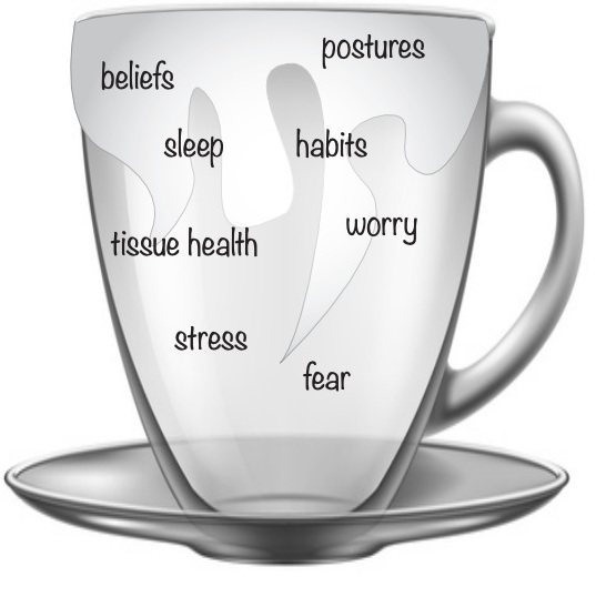
Does this mean posture never matters for pain? No, and most pain science educators will readily admit this. For example, here’s a diagram from Greg Lehman’s workbook, showing the different factors we might hope to modify to help with pain. Note that posture is one of them. But importantly, only one of many.

Screen%2BShot%2B2018-12-24%2Bat%2B2.40.52%2BPM.jpg

4. Biomechanics Are Complex And Not Easy To “Correct.”

Pain science does not overrule the basic laws of physics, and therefore it is obvious that moving with bad technique under heavy load can cause serious injury. But the question remains whether we are likely to get chronic pain from every day habitual movement patterns in low load activities like sitting, standing, breathing, bending, or reaching.  Do these cause repetitive stress and injury, or do we simply adapt to handle the stresses we encounter regularly, just as we do with exercise stress?

This is best answered by looking at a substantial body of research that is similar to the research on posture discussed above. It shows:

  1. Movement patterns that are commonly alleged to be dysfunctional are not correlated with pain or increased injury risk;

  2. Treatments aimed at correcting specific dysfunctions often work no better than general exercise; and

  3. Corrective methods may create good results even when no “correction” occurs, suggesting these methods work by some other mechanism.

For example, general exercise works just as well as stabilization or motor control exercise for low back pain. (Smith 2014; Ferreira 2007; Saragiotto 2016.) This is true even when the treatment involves an attempt to correct a specifically diagnosed movement “impairment.” (Riley 2018, Dillen 2016, Azevedo 2018).

There is a similar pattern for shoulders. General stretching and strengthening works just as well as motor control exercise to correct scapular “dyskinesia.” Further, people improve on motor control programs even when movement patterns stay the same, suggesting the active ingredient is simply exercise, and not a correction of any defect in coordination (See Camargo 2015, McClure 2005, Timmons 2012, Ratcliffe 2014, Struyf 2013). For example, Struyf 2013 concludes that most scapular movement patterns alleged to be dysfunctional probably represent “normal movement variability.”

For knee pain, it seems that the most effective treatment is simply strengthening the muscles around the hip and knee, even when this is done through allegedly “non-functional” exercises with knee extension machines (Willy 2016, Rabelo 2018).

Although Seedman talks at length about the importance of correcting defective movement patterns, he does not specify any such patterns in his article, or how to assess and correct them. He makes vague references to the importance of strong glutes and core muscles for back pain but does not cite any studies on this issue. In fact, core strengthening works no better on average than general exercise for back pain (Lederman). Further, the idea that inactive glutes cause pain is not well-supported by research, which has often shown that pain is associated with more glute activity not less (Kim 2014, Lehman 2006, Suehiro 2015, Dwyer 2013).

The lesson here is that the connection between movement and pain is complex. There’s no doubt that exercise can reduce pain, improve function, and help to prevent injury. But we should be very skeptical about claims concerning the assessment and correction of ”dysfunctions” and their relevance for pain in everyday movements (Tuminello 2017).

5. Inflammation

Seedman suggests that pain scientists ignore the role of inflammation in pain. I have no idea what he might mean here. The role of inflammation in sensitizing nociceptors is one the most basic facts about pain physiology, and also illustrates a key principle of pain education – pain sensitivity can change even as tissue damage stays constant.

Seedman notes that high levels of chronic inflammation are associated with bad health, pain, and reduced function in gait pattern, walking speed, strength, stability, and mobility.  This is in no way surprising. If someone is in poor health and has high levels of inflammation, we would expect that they won’t be moving well or feeling well. Seedman’s evidence demonstrates this obvious connection, but it doesn’t show that the inflammation is caused by poor posture or movement patterns. If it was, then research would easily find correlations between posture and pain, but it doesn’t.

Conclusion: The Truth About Pain Matters

I think getting this information right is incredibly important. Chronic pain is one of the biggest health problems in the world. We have no magic bullets to cure it, but the best hope for progress lies with understanding it better through science and getting people moving. Personal trainers are in an excellent position to do both. There is still much we don’t know, and healthy criticism is needed to move the ball forward. But we know enough to take action – there is a huge gap between common practice and well-established evidence, and that needs to be closed.

Thanks for reading this far! Please share this article with anyone who has questions about pain science. Or point out mistakes if you see them. Please also remember that debate is far more productive when it is respectful and polite.

References (in rough order of appearance.)

O’Sullivan, P. B., Caneiro, J. P., O’Keeffe, M., Smith, A., Dankaerts, W., Fersum, K., & O’Sullivan, K. (2018). Cognitive Functional Therapy: An Integrated Behavioral Approach for the Targeted Management of Disabling Low Back Pain. Physical Therapy, 98(5), 408–423.

Melzack, R. (2010). Pain and the neuromatrix in the brain. Journal of Dental Education, 65(12), 1378–1382.

Brinjikji, W., F. E. Diehn, J. G. Jarvik, C. M. Carr, D. F. Kallmes, M. H. Murad, and P. H. Luetmer. 2015. “MRI Findings of Disc Degeneration Are More Prevalent in Adults with Low Back Pain than in Asymptomatic Controls: A Systematic Review and Meta-Analysis.” American Journal of Neuroradiology 36 (12).

Lederman, E. (2011). The fall of the postural-structural-biomechanical model in manual and physical therapies: Exemplified by lower back pain. Journal of Bodywork and Movement Therapies, 15(2), 131–138.

Foster, N. E., Anema, J. R., Cherkin, D., Chou, R., Cohen, S. P., Gross, D. P., … Woolf, A. (2018). Prevention and treatment of low back pain: evidence, challenges, and promising directions. The Lancet, 391(10137), 2368–2383;

Riley, S. P., Swanson, B. T., & Dyer, E. (2018). Are movement-based classification systems more effective than therapeutic exercise or guideline based care in improving outcomes for patients with chronic low back pain? A systematic review. Journal of Manual & Manipulative Therapy, 1–10.

O’ Sullivan, P., & Caneiro, J. P. (2016). Unraveling the Complexity of Low Back Pain. J Orthop Sports Phys Ther 2016;46(11):932-937.

Smith, B. E., Littlewood, C., & May, S. (2014). An update of stabilisation exercises for low back pain: A systematic review with meta-analysis. BMC Musculoskeletal Disorders, 15(1);

Ferreira, M. L., Ferreira, P. H., Latimer, J., Herbert, R. D., Hodges, P. W., Jennings, M. D., … Refshauge, K. M. (2007). Comparison of general exercise, motor control exercise and spinal manipulative therapy for chronic low back pain: A randomized trial. Pain, 131(1–2), 31–37.

Marcuzzi, A., Wrigley, P. J., Dean, C. M., Graham, P. L., & Hush, J. M. (2018). From acute to persistent low back pain: a longitudinal investigation of somatosensory changes using quantitative sensory testing — an exploratory study. Pain reports, 3, 1–10.

Louw, A., Zimney, K., Puentedura, E. J., & Diener, I. (2016). The efficacy of pain neuroscience education on musculoskeletal pain: A systematic review of the literature. Physiotherapy Theory and Practice, 3985(September).

Borenstein, D G, J W O’Mara, S D Boden, W C Lauerman, A Jacobson, C Platenberg, D Schellinger, and S W Wiesel. 2001. “The Value of Magnetic Resonance Imaging of the Lumbar Spine to Predict Low-Back Pain in Asymptomatic Subjects : A Seven-Year Follow-up Study.” The Journal of Bone and Joint Surgery. American Volume 83–A (9): 1306–11.

Connor, Banks et al. (2003) Magnetic Resonance Imaging of the Asymptomatic Shoulder of Overhead Athletes. A 5-Year Follow-up Study. Am J Sports Med September 2003, Vol. 31, No. 5, 724–727.

Hill, A. L., Aboud, D., Elliott, J., Magnussen, J., Steffens, D., & Hancock, M. (2018). Recover Injury Research Centre and Centre of Research Excellence in Recovery Following Surgical Outcomes Research Centre ( SOuRCe ), Royal Prince Alfred Hospital , Sydney. The Spine Journal.

Tonosu, Juichi, Hiroyuki Oka, Akiro Higashikawa, Hiroshi Okazaki, Sakae Tanaka, and Ko Matsudaira. 2017. “The Associations between Magnetic Resonance Imaging Findings and Low Back Pain: A 10-Year Longitudinal Analysis,” 1–10.

Grundy, P F, and C J Roberts. 1984. “Does Unequal Leg Length Cause Back Pain? A Case-Control Study.” Lancet (London, England) 2 (8397): 256–58.

Pope, M H, T Bevins, D G Wilder, and J W Frymoyer. 1985. “The Relationship between Anthropometric, Postural, Muscular, and Mobility Characteristics of Males Ages 18-55.” Spine 10 (7): 644–48.

Grob, D., H. Frauenfelder, and A. F. Mannion. 2007. “The Association between Cervical Spine Curvature and Neck Pain.” European Spine Journal 16 (5): 669–78.

Nourbakhsh, M R, and A M Arab. 2002. “Relationship between Mechanical Factors and Incidence of Low Back Pain.” The Journal of Orthopaedic and Sports Physical Therapy 32 (9): 447–60.

Dieck, G S, J L Kelsey, V K Goel, M M Panjabi, S D Walter, and M H Laprade. 1985. “An Epidemiologic Study of the Relationship between Postural Asymmetry in the Teen Years and Subsequent Back and Neck Pain.” Spine 10 (10): 872–77.

Franklin, Mary E., and Teresa Conner-Kerr. 1998. “An Analysis of Posture and Back Pain in the First and Third Trimesters of Pregnancy.” Journal of Orthopaedic & Sports Physical Therapy 28 (3): 133–38.

Barrett, Eva, Mary O’Keeffe, Kieran O’Sullivan, Jeremy Lewis, and Karen McCreesh. 2016. “Is Thoracic Spine Posture Associated with Shoulder Pain, Range of Motion and Function? A Systematic Review.” Manual Therapy 26 (December): 38–46.

Richards, K. V., D. J. Beales, A. J. Smith, P. B. O’Sullivan, and L. M. Straker. 2016. “Neck Posture Clusters and Their Association With Biopsychosocial Factors and Neck Pain in Australian Adolescents.” Physical Therapy 96 (10). Oxford University Press: 1576–87.

Andrade, Nicholas S., Carol M. Ashton, Nelda P. Wray, Curtis Brown, and Viktor Bartanusz. 2015. “Systematic Review of Observational Studies Reveals No Association between Low Back Pain and Lumbar Spondylolysis with or without Isthmic Spondylolisthesis.” European Spine Journal 24 (6). Springer Berlin Heidelberg: 1289– 95.

Ettinger, B, D M Black, L Palermo, M C Nevitt, S Melnikoff, and S R Cummings. 1994. “Kyphosis in Older Women and Its Relation to Back Pain, Disability and Osteopenia: The Study of Osteoporotic Fractures.” Osteoporosis International .4 (1): 55–60.

Damasceno, Gerson Moreira, Arthur Sá Ferreira, Leandro Alberto Calazans Nogueira, Felipe José Jandre Reis, Igor Caio Santana Andrade, and Ney Meziat-Filho. 2018. “Text Neck and Neck Pain in 18–21-Year-Old Young Adults.” European Spine Journal 27 (6).

Campos, Tarcisio F de, Chris G Maher, Daniel Steffens, Joel T Fuller, and Mark J Hancock. 2018. “Exercise Programs May Be Effective in Preventing a New Episode of Neck Pain: A Systematic Review and Meta-Analysis.” Journal of Physiotherapy 64 (3). Netherlands: 159–65.

Chaléat-Valayer, Emmanuelle, Jean-Marc Mac-Thiong, Jérôme Paquet, Eric Berthonnaud, Fabienne Siani, and Pierre Roussouly. 2011. “Sagittal Spino-Pelvic Alignment in Chronic Low Back Pain.” European Spine Journal 20 (S5): 634–40.

Smith, Anne, Peter OʼSullivan, and Leon Straker. 2008. “Classification of Sagittal Thoraco-Lumbo-Pelvic Alignment of the Adolescent Spine in Standing and Its Relationship to Low Back Pain.” Spine 33 (19): 2101–7.

Christensen, Sanne Toftgaard, and Jan Hartvigsen. 2008. “Spinal Curves and Health: A Systematic Critical Review of the Epidemiological Literature Dealing With Associations Between Sagittal Spinal Curves and Health.” Journal of Manipulative and Physiological Therapeutics 31 (9): 690–714.

Théroux, Jean, Sylvie Le May, Carole Fortin, and Hubert Labelle. 2015. “Prevalence and Management of Back Pain in Adolescent Idiopathic Scoliosis Patients: A Retrospective Study.” Pain Research & Management : The Journal of the Canadian Pain Society 20 (3). Pulsus Group Inc: 153–57.

Hartvigsen, Jan, Charlotte Leboeuf-Yde, Svend Lings, and Elisabeth H Corder. 2002. “[Does Sitting at Work Cause Low Back Pain?].” Ugeskrift for Laeger 164 (6): 759–61.

Wai, Eugene K., Darren M. Roffey, Paul Bishop, Brian K. Kwon, and Simon Dagenais. 2010. “Causal Assessment of Occupational Lifting and Low Back Pain: Results of a Systematic Review.” The Spine Journal 10 (6): 554–66.

Kwon, B. K., D. M. Roffey, P. B. Bishop, S. Dagenais, and E. K. Wai. 2011. “Systematic Review: Occupational Physical Activity and Low Back Pain.” Occupational Medicine 61 (8): 541–48.

Waddell, G., Burton, A.K., Mar 2001. Occupational health guide- lines for the management of low back pain at work: evidence review. Occup. Med. (Lond) 51 (2), 124e135 (Review).

Shiri, Rahman, Jaro Karppinen, Päivi Leino-Arjas, Svetlana Solovieva, and Eira Viikari-Juntura. 2010. “The Association between Smoking and Low Back Pain: A Meta- Analysis.” The American Journal of Medicine 123 (1): 87.e7-87.e35.

Hoogendoorn, W E, M N van Poppel, P M Bongers, B W Koes, and L M Bouter. 2000. “Systematic Review of Psychosocial Factors at Work and Private Life as Risk Factors for Back Pain.” Spine 25 (16): 2114–25.

Hodges, Paul W., G. Lorimer Moseley, Anna Gabrielsson, and Simon C. Gandevia. 2003. “Experimental Muscle Pain Changes Feedforward Postural Responses of the Trunk Muscles.” Experimental Brain Research 151 (2): 262–71.

Saragiotto, Bruno T, Christopher G Maher, Tie P Yamato, Leonardo O P Costa, Luciola C Menezes Costa, Raymond W J G Ostelo, and Luciana G Macedo. 2016. “Motor Control Exercise for Chronic Non-Specific Low-Back Pain.” The Cochrane Database of Systematic Reviews, no. 1 (January). England: CD012004.

Riley, S. P., Swanson, B. T., & Dyer, E. (2018). Are movement-based classification systems more effective than therapeutic exercise or guideline based care in improving outcomes for patients with chronic low back pain? A systematic review. Journal of Manual & Manipulative Therapy, 1–10.

Azevedo, Daniel Camara, Paulo Henrique Ferreira, Henrique de Oliveira Santos, Daniel Ribeiro Oliveira, Joao Victor Leite de Souza, and Leonardo Oliveira Pena Costa. 2018. “Movement System Impairment-Based Classification Treatment Versus General Exercises for Chronic Low Back Pain: Randomized Controlled Trial.” Physical Therapy 98 (1). United States: 28–39.

Dillen, L., et al. 2017. Efficacy of classification-specific treatment and adherence on outcomes in people with chronic low back pain. A one-year follow-up, prospective, randomized, controlled clinical trial. Man Ther. 2016 August ; 24: 52–64

Timmons, Mark K, Chuck A Thigpen, Amee L Seitz, Andrew R Karduna, Brent L Arnold, and Lori A Michener. 2012. “Scapular Kinematics and Subacromial-Impingement Syndrome: A Meta-Analysis.” Journal of Sport Rehabilitation 21 (4). United States: 354–70.

Ratcliffe, Elizabeth, Sharon Pickering, Sionnadh McLean, and Jeremy Lewis. 2014. “Is There a Relationship between Subacromial Impingement Syndrome and Scapular Orientation? A Systematic Review.” British Journal of Sports Medicine 48 (16): 1251–56.

Struyf, F., J. Nijs, S. Mollekens, I. Jeurissen, S. Truijen, S. Mottram, and R. Meeusen. 2013. “Scapular-Focused Treatment in Patients with Shoulder Impingement Syndrome: A Randomized Clinical Trial.” Clinical Rheumatology 32 (1). Springer-Verlag: 73–85.

Camargo, Paula R, Francisco Alburquerque-Sendin, Mariana A Avila, Melina N Haik, Amilton Vieira, and Tania F Salvini. 2015. “Effects of Stretching and Strengthening Exercises, With and Without Manual Therapy, on Scapular Kinematics, Function, and Pain in Individuals With Shoulder Impingement: A Randomized Controlled Trial.” The Journal of Orthopaedic and Sports Physical Therapy 45 (12). United States: 984–97.

McClure, Philip W, Jason Bialker, Nancy Neff, Gerald Williams, and Andrew Karduna. 2004. “Shoulder Function and 3-Dimensional Kinematics in People with Shoulder Impingement Syndrome before and after a 6-Week Exercise Program.” Physical Therapy 84 (9). United States: 832–48.

Willy, R. W., & Meira, E. P. (2016). Current Concepts in Biomechanical Interventions for Patellofemoral Pain. International Journal of Sports Physical Therapy, 11(6), 877.

Rabelo, N. D. D. A., & Lucareli, P. R. G. (2018). Do hip muscle weakness and dynamic knee valgus matter for the clinical evaluation and decision-making process in patients with patellofemoral pain? Brazilian Journal of Physical Therapy, 22(2), 105–109.

Kim, Ji-Won, Oh-Yun Kwon, Tae-Ho Kim, Duk-Hyun An, and Jae-Seop Oh. 2014. “Effects of External Pelvic Compression on Trunk and Hip Muscle EMG Activity during Prone Hip Extension in Females with Chronic Low Back Pain.” Manual Therapy 19 (5). Scotland: 467–71.

Lehman, Gregory J. 2006. “Trunk and Hip Muscle Recruitment Patterns during the Prone Leg Extension Following a Lateral Ankle Sprain: A Prospective Case Study Pre and Post Injury.” Chiropractic & Osteopathy 14 (February). England: 4.

Suehiro, Tadanobu, Masatoshi Mizutani, Hiroshi Ishida, Kenichi Kobara, Hiroshi Osaka, and Susumu Watanabe. 2015. “Individuals with Chronic Low Back Pain Demonstrate Delayed Onset of the Back Muscle Activity during Prone Hip Extension.” Journal of Electromyography and Kinesiology : Official Journal of the International Society of Electrophysiological Kinesiology 25 (4). England: 675–80.

Dwyer, Maureen K, Kelly Stafford, Carl G Mattacola, Timothy L Uhl, and Mauro Giordani. 2013. “Comparison of Gluteus Medius Muscle Activity during Functional Tasks in Individuals with and without Osteoarthritis of the Hip Joint.” Clinical Biomechanics (Bristol, Avon) 28 (7). England: 757–61.

Tumminello, N., Silvernail, J., & Cormack, B. (2017). The corrective exercise trap. Personal Training Quarterly, 4(1), 6–15.

Painful Exercise In Rehab – Yes, No, Or A Lot To Think About

Whether rehab exercises should be painful or not has become a recent topic of much discussion. As we have started to value the role of (optimal) loading in rehab, and that we can get patients back to moving and loading pretty early in the rehab process, we have also got to appreciate that this is unlikely to happen without experiencing any pain.

If you have any type of understanding about pain then the simple question of “should we use painful exercise?” suddenly might not look so simple!

Rather than a simple yes or no, there are a bunch of questions to be answered, such as how much pain is OK? Who may or may not benefit? And what is the best way to manage the process? Especially if all does not go to plan!

Research

From a data standpoint it would be great if we had have something that we can use to guide us here. Low and behold we do, and its open access.

Smith et al looked at this question in 2017.

Should exercises be painful in the management of chronic musculoskeletal pain? A systematic review and meta-analysis

This systematic review and meta analysis looked at 9 trials in which varying degrees of painful exercise were used. They found that painful exercise did NOT result in statistically worse outcomes across a short, medium or long term follow up. There was a small statistical benefit in the short-term for painful exercises as highlighted by the standardised mean difference of around 0.2 (SMD = effect size for a meta analysis). So from a research standpoint this is positive news.

One thing to note here from a clinical standpoint is, that using painful exercise is no GUARANTEE that it will have a positive effect for your patient. All statistical tests only give us the PROBABILITY of an effect, so the likelihood is that it will, but when we look at the confidence intervals (measure of variability) for the mean SMD for painful exercise, for the majority of the studies we can also see that they cross into a negative effect and in some of the studies quite substantially.

The variable effect of exercise on pain (although over a shorter time frame) was also shown by O Neill et al’s recent look at the acute effects of isometrics *Here*. Although, of course, isometric exercise and painful exercise is not the same thing, the point here is the highlighting of the variable effects from exercise. For patients with higher baseline pain isometrics actually increased their pain post exercise and this is the benefit of looking at individual responders within a study not just the group average. Those with lower pain at baseline seemed to receive more analgesia from isometric exercise.

BUT at the very least I think we can say that the likelihood is that it will not have detrimental effect. So as rehab can often be painful we can be reasonably confident but without seeing painful exercise as some sort of fire and forget panacea and also appreciate the individual nature of pain.

We could debate the mechanisms of HOW painful exercise might work from pain habituation to conditioned pain modulation and perceptual mechanisms but in essence we don’t really know at this point in time and this might be different or in different combinations for different people.

Benefits

The benefits can be both physical AND psychological.

Firstly, it allows patients to get in MORE exercise. If they are not being limited by pain (or pain contingent if we are being fancy :) then they can get a greater dosing than if they stopped due to it being painful. It is important to add though that we currently don’t know what optimal dosing is for exercise having an effect on pain!

Secondly, it sends the message that hurt does not equal harm. The problem can be painful and still settle down if managed in the right way. This may give people a new insight into their pain and how they can manage it.

Pain self-efficacy may also be built experientially, so using painful exercise as a tool might be an option to help do this.

Clinical Application

So it’s great to have some objective data on this subject, but as we know getting that into clinical application is not always straightforward.

Firstly don’t be afraid of some pain! The likelihood is that it won’t cause a worse outcome. But as we are dealing with thinking, feeling HUMANS, it is OF COURSE not as simple as this, but more on that later.

One of the issues with always looking to be pain free with exercise or continually treating pain, is what does that say ABOUT pain? On one hand we are telling people hurt does not mean harm but our actions might not be saying the same thing. If pain is OK, if it is normal then in the process of getting it better we might have to endure a little bit of it.

But how much is OK is a key question? Studies that have allowed painful exercise have ranged from not increasing baseline symptoms during or post exercise up to 5/10 on the VAS.

We know the limitations of the VAS as a REALLY subjective scale so perhaps we could introduce the idea of pain being tolerable or intolerable rather than a numerical score.

Screen Shot 2019 01 21 At 13.16.00


Now tolerable might be a bit sore but it does not really get in the way. Intolerable might be thought of as getting in the way of our daily activates, maybe having an effect on our sleep and generally making life a bit miserable. This is going to be different for different folk so hence why the concept of tolerable becomes important.

We would also like to know how the pain is settling down. So pain that is not really calming down after the exercise stimulus or keeps on getting progressively worse over a number of days as the exercises are performed is probably not a great idea. We would like to see a nice stimulus response with only slight increases above baseline (tolerable remember : ) that calms down gradually over 24-36 hours, quite similar to delayed onset muscle soreness (DOMS)

So on the issue of DOMS, it is important to identify if the increase in pain is actually the same as the original symptom. Post exercise soreness is quite normal but for those that are unaccustomed to exercise this might not be recognisable especially for a currently painful area. Defining what is pain and what is soreness might be a good sense making exercise for some patients. Although we don’t know that progressive overload is required with exercise in rehab, it probably ia a good idea, so having some sort of tolerable baseline for patients to progress or regress from is a great idea too. This is also might have an effect their self efficacy using exercise too which, of course, is a bonus.

Pain Is Not The Only Issue

Potentially the most problematic area with painful exercise, and one that is really under discussed, may be more about the psychological aspects that are associated with the pain and how they can affect the person. One of the best prognostic factors for recovery in a number of body areas appears to be pain self efficacy, *here* is a recent paper from Chester et al and also from Foster et al *here*  Pain self efficacy, something I have written about before also *here*, is the ability to carry on normal functioning despite of pain. It is important to note that those with higher pain self efficacy also tend to adhere to exercise better.

So someone’s pain self efficacy is going to be a KEY factor in whether they are able to tolerate having pain and being able to carry on functioning which maybe a fundamental skill at the heart of painful exercise especially if it can take 24-36 hrs to settle.

Predicted outcome is another key prognostic factor and if someone believes that increased pain will result in a negative outcome then this could have a negative effect on the actual outcome.

Jack et al *here* found that the number one reason for poor adherence to exercise programs was that people did not want to make the problem worse. So painful exercise coupled with negative beliefs about pain, low pain self efficacy and a poor predicted outcome may not sit well together.

A qualitative piece “Exploring experiences, barriers, and enablers to home- and class-based exercise in rotator cuff tendinopathy” from Sandford et al also found that a fear of making the issue worse was a key factor in reduced adherence to exercise.

What Can We Do?

Firstly you could screen people for these factors if you feel they are required. We have tools such the Pain Self Efficacy Questionnaire  (PSEQ) and the shorter version the PSEQ-2.

One caveat with using questionnaires, in my opinion : ), is it does allow us to get a score to assess confidence and resilience in managing pain but it perhaps does not tell us about things that are specific to our patients. So being able to weave this into a good subjective that teases these more personal parts of the narrative out is a must.

Also ASK about their expected outcome from the treatment and what would it mean to you if it was painful.

“Do you feel like this exercise will work for you?”

“Do you have any concerns about doing this exercise?”

It might be that someone tells you that they have tried exercises before and they did not work or that they feel it might make the issue worse, especially if it hurts.

It might also be important to discuss with ALL patients what their beliefs are around pain. These might be that pain indicates they are doing more damage to their bodies or they will not be able to work because they have pain or implications for future functioning. It can often be what the pain MEANS to the patient that is the real issue rather than just the sensation of the pain itself.

A key factor to remember is regardless of whether painful exercise is positive or no worse than non painful exercise, a negative belief may stop your patient from exercising

Education

One of the most powerful tools we have at our disposal is EDUCATION, I am not always a huge fan of this term as it can imply a teacher pupil relationship. But in order to provide effective education to people we first have to build rapport and then understand what they may need to know more about.

We can educate on a range of things from the effectiveness of exercise for many issues, that painful exercise is often not detrimental, that pain self efficacy is an important prognostic factor and what pain may actually mean and its relationship with physical damage. It may also be worth talking about the process and what to expect, and often that it is a process of trial and error to get the right level and desired response.

This has to start with understanding the patient, their history and belief structure.

Where You Start Is Not Where You Have To Finish

Do we have to jump straight into painful exercise? I don’t think so. It is not REQUIRED but it is also not harmful.

An important point to make is that for someone with very negative beliefs about pain, reduced pain self efficacy and poor beliefs about the outcome, challenging them right off the bat with painful exercise might not always be the best thing to do. As they build trust in you and confidence in their bodies then pushing into pain progressively might sometimes be a better route.

We know that giving people the option to avoid pain can actually maintain pain avoidance, so whilst not advocating avoidance behaviour confronting these things may take a little time, confidence and education, especially if the person has a lot of negative pain and avoidance based beliefs.

Those that persist in doing painful things that have NOT provide a therapeutic effect may benefit from non painful exercise for a while too. If you feel that pushing into pain has not given the desired response when looking at their history then a break or change in dosage could be just the ticket.

Those with higher levels of pain at baseline on average seem to have worse clinical outcomes so this might also lead me to reason that maintaining this level of pain through exercise might not always be a good idea.

There are no real hard and fast rules here only individuals and individual considerations.

What If It All Goes Wrong???

This is where self efficacy may come into its own. Empowering your patients to self manage maybe valuable in the exercise process.

As we hopefully have told the patient BEFORE they embark on an exercise program I can often be trial and error. Have we enabled our patients to adapt their exercise dosage if it goes a bit wrong? *Here* is a short piece on dosage in rehab

So do your patients know what to look out for in terms of symptoms?

Do they know how to adjust the dosage? This could be in exercise frequency, intensity and time, sets and reps (volume).

Are they able to contact you to ask questions? Do they know this?

I think these are all required aspects of pushing into pain.

A patient I saw last week had religiously persisted at pushing into his painful rehab exercises every 48hrs. He believed if he did what he was told he would get better (the trust!). But after 3 months there was not the desired outcome. In this case he was given no indication what to do if this happened, just blind faith on all sides that he would get better.

Key Points

  • Painful exercise DOES NOT produce worse outcomes from the research we have
  • Research does not guarantee your patients response to painful exercise
  • It is not just about the PAIN sensation! What does the pain mean and how does it affect behaviour such as exercise adherence?
  • Pain self efficacy and predicted outcome are important psychological measures and can be discussed and measured
  • Think about education around the exercise and the pain response
  • It does not need to start with painful exercise!
  • Empower your patients to self manage


Why Weak Glutes Aren’t A Reliable Predictor Of Low Back Pain

It seems we are constantly trying to find causes for low back pain with our patients.

Quite often we are caught up looking for biomechanical issues (which there often are) and avoiding other things that could influence a person’s pain.

One such instance is the possibility that weak glutes are a contributing factor.

I recall seeing one of my instructors demonstrate this (what I perceived to be) complicated movement test where my patient was lying prone and having them extend their leg. There was a specific pattern where the muscles in the low back down through the glutes and hamstrings were supposed to move.

This would be followed up with the comment: “your glutes aren’t firing”!

While I watched this all I could think was…”there’s no way I’m going to remember what order all of this goes in”!

But, when done with confidence and a really good explanation, it seemed to make sense to me, and the patient.

So, I worked through the low back, glutes, and hamstrings, and the patient felt better after the treatment.

I proceeded to pat myself on the back for a job well done!

However, the next time a patient came in for what I thought was the same issue, I couldn’t remember this specific pattern of movement and what it was supposed to tell me! Why couldn’t I get this!?

Well, in reality, I really didn’t have to and here’s why.

Limitations With Testing

There are a couple of tests used to see if the glutes are a contributing factor to low back pain, so let’s look at both of them.

The prone leg extension test1 is pretty commonly used to test for low back pain and lumbopelvic function (this is the one my instructor was demonstrating to me).

The pattern of movement you’re supposed to see is (if you were testing the right side) right glute max, right hamstring, left lumbar erector spinae, right lumbar erector spinae, left thoracolumbar erector spinae, then the right thoracolumbar erector spinae. A delay in glute max recruitment is supposed to show a dysfunctional pattern of movement.

There is a whole list of other patterns that are supposed to mean other things, but it’s a long list and more than I want to get into for the purposes of this post.

A studyshowed there was no consistent order of activation during the test and glute max was the last muscle to become activate with time ranging from 0.07 to 0.676 seconds in delay.

Now, these tests were done with proper EMG testing, so I have to wonder…how would we ever be able to properly assess this just using touch and vision!?

Another test we were taught was “Trendelenburg”, or “SLS” to assess for glute weakness possibly contributing to back pain. 

With this test, a practitioner stands behind the patient and has them raise one leg so the hip is between 60°-90° of flexion. A positive test shows lateral pelvic tilt on the stance leg, which is supposed to represent glute weakness on that side.

One study 2 compared the differences between those with chronic low back pain vs. a control group using Trendelenberg. It actually showed the number of positive tests was no different between the groups. There was also no difference in glute med strength between those who scored a positive and negative test.

Since there was an equal number of positive and negative tests between both groups it showed this test is unreliable to differentiate between those with chronic low back pain and those without, nor was it reliable for demonstrating a difference in glute med strength.

Another study 3 actually injected a superior gluteal nerve block to see if there were any alterations in different movement variables. They found after the injection there was no difference in contralateral pelvic drop, hip adduction, or hip abduction due to reduced strength of the glutes post-injection.

The researchers found this surprising as an impaired gluteal nerve has always been associated with the SLS test.

So, in other words, lateral pelvic drop is not associated with weak glute med muscles in our patients who are dealing with chronic low back pain. It was even suggested that glute med strength of less than 10% of body weight is required to actually get a positive SLS.

The suggestion was also made that in order to maintain your stance during this test it is because of all muscles attaching to the greater trochanter working together combined with the mechanical force of the iliotibial band. This shows a positive SLS is more likely a global issue rather than a specific spine and pelvis issue.

In light of their findings they showed how this test could not distinguish between who was experiencing low back pain and who wasn’t, nor could it identify those who had weak glutes, and in fact demonstrated the test was probably due to some other issue altogether.

Another study 4 showed the presence of low back pain was a combination of higher BMI, signs of hip abductor problems, significant glute med weakness, gluteal tenderness, and a positive SLS (but the strength testing was admittedly not reliable in the study). However, this study showed something else that is probably far more significant which we will look into next.

It was unclear if glute med muscle weakness was the cause of the low back pain…or just a consequence of it.

Muscle Activation, Weakness, Or Protection?

Quite often when discussing this kind of thing and it’s relation to low back pain, the discussion can revolve around not only issues with the glutes, but also hamstring length and activation patterns between the two.

Inevitably when discussing “weak glutes” it’s due to an S.I. joint “dysfunction”, or this dysfunction is causing the weak glutes. But really, what is dysfunctional with the S.I. joint in this case?

They 5 described S.I joint dysfunction as; low back pain below L5, pain over the posterior aspect of SI joint around PSIS and buttock with or without above the knee leg pain. They used a combination of three SI joint provocation tests (posterior shear, compression, distraction, and sacral thrust) that showed good sensitivity and specificity (although the sensitivity rating was much higher).

When we look at the discussion around which muscles are “activating” with certain movements it is usually centered around the context of the pain-spasm-pain model which suggests pain results in increased muscle activity, in turn creating more pain.4

Or, the pain adaptation model which suggests pain reduces activation of muscles when active as agonists and increases activation of muscles when active as antagonists.They say this will reduce movement and ROM which would, in turn, prevent mechanical pain and further damage to the tissues.

The funny thing is that while both of these models are trying to prove the same thing, they end up contradicting each other quite a bit and neither of these ideas can predict how back pain will affect muscle activation.

There is lots of discussion around both increased and decreased muscle activation due to pain which results in disrupting motor control. However, this systematic review 6 argues that increases in muscle activation is an adaptation that occurs for various reasons, and those changes are mainly to avoid harmful stress placed on already injured structures, and to increase stability around the spine.

In each case, they looked at any changes that were task-dependent, related to an individual problem, and highly variable between each patient. This also shows those changes are functional since they are trying to reduce unpleasant stress and provide stabilization to the spine.

One common thread through most of the research cited is glute weakness in relation to back pain. However, it’s a bit of a chicken or the egg, is the weakness a result of reduced use because of pain, or did the weakness cause the pain? I’d venture to say it’s due to the reduced use.

While this can all sound maybe a little complicated and like word salad, when we look at everything we know about pain, what is this actually telling us?

One thing we know for sure that pain is ALWAYS a protection mechanism. So, if something in our body has a decreased ROM for a period of time in order to prevent stress in a sensitive area, this is just simply a way to protect that area. If those altered movements are also providing extra support to stabilize something, it’s actually doing this to protect the area! With this understanding we have to ask, does it really matter what order a muscle fires in (especially because we’d never be able to accurately assess this with just the human eye)? What we should be concerned with is calming those sensitized areas down, reassuring the patient they aren’t broken, and educating them as to why they’ll be okay. Then get them moving in pain-free full ranges again and help strengthen the area, build up that resilience!


References

  1. Lehman GJ, Lennon D, Tresidder B, Rayfield B, Poschar M. Muscle recruitment patterns during the prone leg extension. BMC Musculoskeletal Disorders. 2004 Dec 1;5(1):3.
  2. Penney T, Ploughman M, Austin MW, Behm DG, Byrne JM. Determining the Activation of Gluteus Medius and the Validity of the Single Leg Stance Test in Chronic, Nonspecific Low Back Pain. Archives of Physical Medicine & Rehabilitation [Internet]. 2014 Oct [cited 2020 Apr 17];95(10):1969–76. Available from: https://search.ebscohost.com/login.aspx?direct=true&db=s3h&AN=98597253&site=ehost-live
  3. Pohl MB, Kendall KD, Patel C, Wiley JP, Emery C, Ferber R. Experimentally reduced hip-abductor muscle strength and frontal-plane biomechanics during walking. Journal of athletic training. 2015 Apr;50(4):385-91.
  4. Cooper NA, Scavo KM, Strickland KJ, Tipayamongkol N, Nicholson JD, Bewyer DC, Sluka KA. Prevalence of gluteus medius weakness in people with chronic low back pain compared to healthy controls. European Spine Journal. 2016 Apr 1;25(4):1258-65.
  5. MassoudArab A, RezaNourbakhsh M, Mohammadifar A. The relationship between hamstring length and gluteal muscle strength in individuals with sacroiliac joint dysfunction. Journal of Manual & Manipulative Therapy. 2011 Feb 1;19(1):5-10.
  6. van Dieën JH, Selen LP, Cholewicki J. Trunk muscle activation in low-back pain patients, an analysis of the literature. Journal of electromyography and kinesiology. 2003 Aug 1;13(4):333-51.


Articles Of The Week April 19, 2020


How often do you have a patient come in who is dealing with pelvic pain? I haven’t seen it much in my practice but I think this is also partially because it’s not common for a Massage Therapist to treat this area. Well, perhaps it’s time we change that.

“Treatment of Pelvic and Abdominal Pain for the RMT” – Jocelyn Kirton

We all know I have mad respect for Todd Hargrove, so when he puts on a master class about pain, movement, and play (while also doing it for free), I think we should all take advantage!

“Pain, Movement, And Play: Common Sense For Complex Problems” – Todd Hargrove

I haven’t had a chance to do this course myself but it was recommended by someone I have HUGE respect for, so it must be good. Another great way to get some learning done during the pandemic.

“Exercise And Physical Activity For Knee Osteoarthritis” – Rana Hinman

Still not sure where to start learning about pain? This is a phenomenal resource put together on some of the top pain resources available.

“Pain Resources: A Deeper Dive” – Rebecca Hall

We all know how important exercise is. As this review points out, it’s actually very important to help decelerate cognitive decline. Although exercise might not enhance cognitive function, it does effectively decelerate the decline in cognitive function.

“The Effect of Physical Exercise on Cognitive Functioning in Individuals with MCI and Dementia” – Scott Buxton


Looking at Pain From Different “Levels”


Pain can be a complex phenomenon, meaning that many different factors might contribute to pain, and that these factors can be interwoven in a way that makes it very hard to separate one from the other, either for purposes of explaining pain or intervening to treat it in some way.

This is a major theme of my recent book Playing With Movement, where I argue that complexity science has many concepts that are useful in understanding movement and pain.

One of these concepts is that complex systems are often nested. That means the system as a whole is composed of smaller subsystems, which are also composed of smaller subsystems and so forth.


Example of pain complexity

Where is the pain?

For example, people are made up of organ systems (like the nervous system or musculoskeletal system), which are in turn composed of organs (like the brain and spinal cord, muscles, and tendons), which are composed of cells (like nerve cells and muscle cells) and so forth. Further, people are parts of larger systems like families and communities and economies. This is interesting because each nested system provides a different level from which we can attempt to explain and treat pain.
Here’s a diagram to illustrate:
Screen Shot 2018-05-11 at 7.17.42 PM.png

At the “lower” levels, you can analyze the health status of cells and organs like muscles, tendons, discs or nerves. For example, maybe your foot hurts because of a stress fracture. This is where you can find “issues in the tissues”, which is where traditional pain treatment has focused most of its attention. This is often called the “biomedical approach” or the “bio” part of the biopsychosocial model. You find the structure that is damaged and work to repair it.

At the “higher” levels of analysis, such as the person or the environment, you are looking at more complex phenomena – the role of thoughts, emotions, or social relationships. These are the “psychosocial” issues that are known to have very important effects on chronic pain.

Problems in these areas are often relatively subtle, more about dysregulation or imbalance than something being broken or injured. These issues are also invisible if you look for them at a lower level. For example, you can’t see catastrophising by assessing a foot – you need to talk to a person.

Fields Of Study

There are many different formal disciplines you could study to get a better understanding at each level. Note that they are very different from one another, and very few people will have significant knowledge at more than one level.

Screen Shot 2018-05-11 at 7.19.35 PM.png

At the lower levels, you could study biomechanics, exercise physiology or neurodynamics. Each would give you a better understanding of how physical structures in the body respond to stress – either by breaking down and getting injured or adapting to get stronger.

You could move up a level to study the behavior of a larger system like the nervous system, immune system or endocrine system. This would help you see that pain functions like an alarm. The nervous, immune and endocrine systems help set the sensitivity of the alarm, and determine the kinds of events that cause it to go off. “Pain science” is mostly education in the basic physiology of these systems as they relate to pain. 

We can move up another level to the “person”, where we are studying the role of cognitions and emotions in pain. This is the realm of psychology, the relevance of which should be obvious – pain is a psychological event.

Psychological concepts can be very useful in understanding why movement and physical activity help with pain. For example, cognitive behavioral therapy can explain how exercise might extinguish fears or expectancies that contribute to pain. In many cases, this perspective is more helpful in choosing an exercise program than one focused on “lower-level” concerns about muscle groups, reps, and sets.   

You could move yet higher to study the role of social and economic systems. Many social critics argue that the real pathologies causing a wide variety of chronic diseases – including drug addiction, anxiety, depression, and even chronic pain – live more at the level of society than the individual. For example, low socioeconomic status is a big predictor of chronic pain. Most readers of this blog are not active in trying to solve problems at this level but are quite aware that they have a big impact on clinical outcomes.

Comparing The Different Levels

The terms “high level” and “low level” don’t reflect any value judgment. They merely indicate different perspectives: one is taking a “micro” view of relatively small and simple things like tendons or muscles, and the other is taking a “macro” or big picture look at larger complex things like nervous systems and emotions.

In general, if you’re moving down levels in your effort to explain some problem, you could call that “reductionist.” And if you’re moving up, that might be called a more “holistic” or “systems thinking” approach.

Screen Shot 2018-05-11 at 7.37.06 PM.png
Again, there is not necessarily any right or wrong here – the right level depends on the context.  Some problems with pain, especially those related to acute injuries, benefit from a lower-level approach – strengthen this, stretch that, do X sets of Y reps for Z weeks and then you will be fixed. Other pain problems can never really be “fixed” and may be hard to manage even with a whole team of psychotherapists, social workers, and attorneys.
Although each end of the spectrum has its costs and benefits, there is no doubt that until very recently, manual and movement therapists have spent way too much time at the lower levels, looking for issues in the tissues with their microscopes, while ignoring some very real big picture human issues sitting right in front of them. If the “pain science revolution” means anything, it is trying to improve basic literacy at the higher levels. “Playing with movement” means exploring a problem from as many different levels and perspectives as possible. To put it simply, finding physical activities that are challenging, meaningful and variable will go along way toward engaging us on almost any level that matters.